. 2003 Nov;52(11):693-8.
doi: 10.1007/s00262-003-0407-6. Epub 2003 Jun 24.

L-ascorbic acid (vitamin C) induces the apoptosis of B16 murine melanoma cells via a caspase-8-independent pathway

Affiliations

L-ascorbic acid (vitamin C) induces the apoptosis of B16 murine melanoma cells via a caspase-8-independent pathway

Jae Seung Kang et al. Cancer Immunol Immunother. 2003 Nov.

Abstract

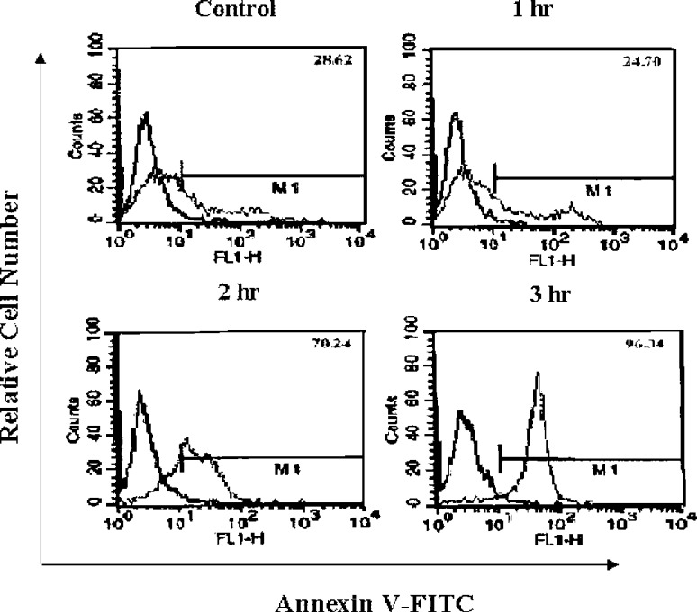
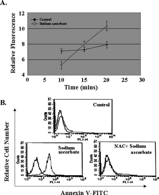
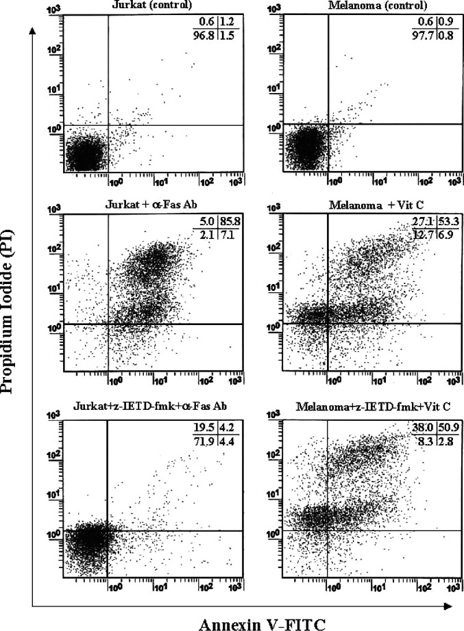
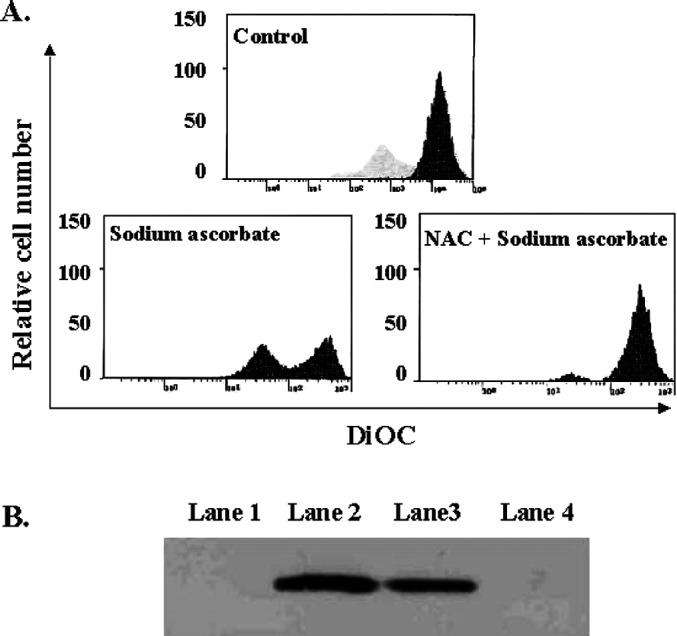
L-ascorbic acid (vitamin C) has been reported to play a role in the treatment and prevention of cancer. However, its specific mechanistic pathways remain obscure. This study was carried out to identify the sodium ascorbate-induced apoptotic pathway in B16F10 murine melanoma cells. Sodium ascorbate was found to induce the apoptosis of B16F10 murine melanoma in a time- and dose-dependent manner, and this was prevented by pretreatment with N-acetyl- L-cysteine (NAC), a well-known antioxidant. In fact, sodium ascorbate-treated B16F10 melanoma cells showed increased intracellular reactive oxygen species generation (ROS) levels. These results indicate that sodium ascorbate induced apoptosis in B16F10 murine melanoma cells by acting as a prooxidant. We examined the involvement of caspase-8 using a specific caspase-8 inhibitor (z-IETD-fmk) on the sodium ascorbate-induced apoptotic pathway. Cell death was found not to be inhibited by z-IETD-fmk treatment, indicating that sodium ascorbate-induced apoptosis is not mediated by caspase-8. In addition, we detected a reduction in the mitochondrial membrane potential during apoptosis and confirmed cytochrome-c release from mitochondria by immunoblotting. Taken together, it appears that the induction of a prooxidant state by sodium ascorbate and a subsequent reduction in mitochondrial membrane potential are involved in the apoptotic pathway of B16F10 murine melanoma cells, and that this occurs in a caspase-8-independent manner.


Figures

Fig. 1.
Fig. 1.
Kinetics of sodium ascorbate–induced apoptosis. Cells were incubated with media (control) or sodium ascorbate (10 mM) for various time periods as indicated, at 37°C in a 5% CO2 humidified incubator. After incubation, cells were harvested and apoptosis was detected by Annexin V-FITC analysis. Results are representative of more than five experiments
Fig. 2A, B.
Fig. 2A, B.
Effect of sodium ascorbate on apoptosis as a prooxidant. A Effect of sodium ascorbate on ROS levels of B16F10 melanoma cells. Cells (2×10⁴ cells/well) were placed in wells of a 96-well microtiter plate followed by an addition of 50-μM 2', 7'-dichlorofluorescein diacetate at 37°C in a 5% CO2 humidified incubator, and then analyzed with a Cytofluor 2350 plate reader with excitation at 485 nm and emission at 530 nm. Results are representative of more than five experiments, each performed in triplicate. The data are shown as the mean ± SD. B Effect of the antioxidant, N-acetyl-l-cysteine on sodium ascorbate–induced apoptosis. B16F10 melanoma cells were incubated with medium (control), sodium ascorbate (10 mM), or sodium ascorbate/N-acetyl-l-cysteine (10 mM) for 4 h. After incubation, cells were collected and stained with Annexin V-FITC to detect apoptosis of melanoma cells. Results are representative of more than five experiments
Fig. 3A–F.
Fig. 3A–F.
Caspase-8 is not involved in the sodium ascorbate–induced apoptosis. Jurkat and B16F10 melanoma cells (1×10⁶ cells/ml) were pretreated for 30 min at 37 C with z-IETD-fmk (20 mM). Following this incubation, targets were treated with antihuman Fas Ab (2 ng/ml) for 24 h at 37°C and 10 mM of Sodium ascorbate for 4 h at 37°C, and then apoptosis was determined using Annexin V-FITC apoptosis detection kit. A Jurkat cells (control), B Jurkat cells + anti-Fas Ab (2 ng/ml), C Jurkat cells + z-IETD-fmk (20 mM) + anti-Fas Ab (2 ng/ml), D B16F10 melanoma cells (control), E B16F10 melanoma cells + sodium ascorbate (10 mM), F B16F10 melanoma cells + z-IETD-fmk (20 mM) + sodium ascorbate (10 mM). Results are representative of more than five experiments
Fig. 4.
Fig. 4.
Sodium ascorbate induces apoptosis on B16 murine melanoma cells via the decrease of mitochondrial potential and the release of cytochrom C. A. Measurement of mitochondrial membrane potential: 10 mM of N-acetyl-l-cysteine pretreated or untreated B16F10 melanoma cells were cultured with 10 mM of sodium ascorbate and then stained with DiOC6. Results are representative of three experiments. B Cytochrome-c release induced by sodium ascorbate; Jurkats were incubated without (Lane 1) or with (Lane 2) anti-Fas Ab for 24 h at 37°C. B16F10 murine melanoma cells were incubated with (Lane 3) or without (Lane 4) 10 mM of sodium ascorbate for 4 h at 37°C. Cells were washed in PBS and resuspended in digitonin lysis buffer for 5 min on ice followed by centrifugation. Fifty microliters of cytosolic proteins in SDS-loading buffer were loaded onto 15% polyacrylamide gels. Following SDS-PAGE, proteins were transferred to nitrocellulose membranes and probed with a monoclonal anti-cytochrome-c Ab (1 μg/ml). After the addition of a biotin conjugated-antimouse Ab (1:5,000), proteins were detected using ECL. Results are representative of more than five experiments

References

    1. Afansev FEBS Letter. 1986;194:347. doi: 10.1016/0014-5793(86)80114-4. - DOI
    1. Bram Nature. 1980;284:629. - PubMed
    1. De Anticancer Res. 1990;10:391. - PubMed
    1. De Anticancer Res. 1990;10:1029. - PubMed
    1. Du Cell. 2000;102:33. - PubMed

Publication types