Comparative Study
. 2003 Jul;85(1):300-12.
doi: 10.1016/S0006-3495(03)74475-0.

The pore helix is involved in stabilizing the open state of inwardly rectifying K+ channels

Affiliations
Comparative Study

The pore helix is involved in stabilizing the open state of inwardly rectifying K+ channels

Noga Alagem et al. Biophys J. 2003 Jul.

Abstract

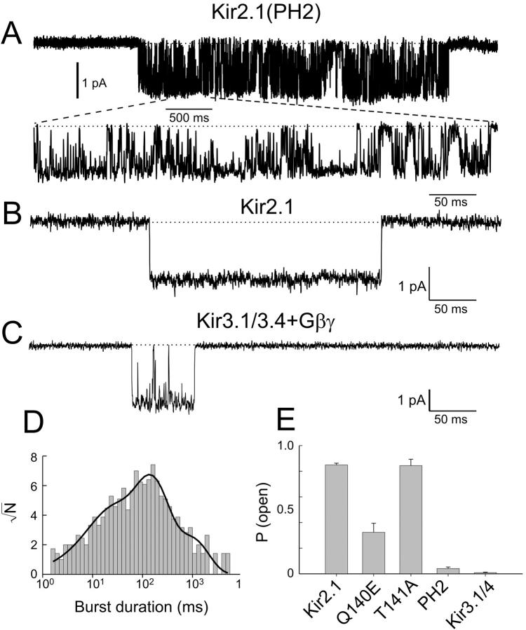
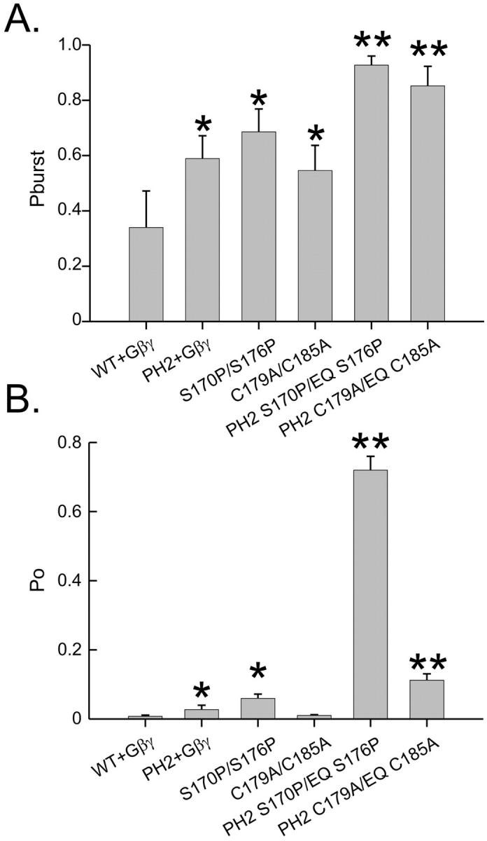
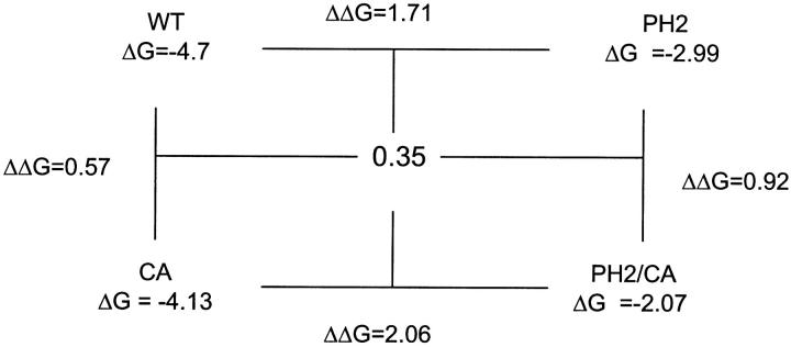
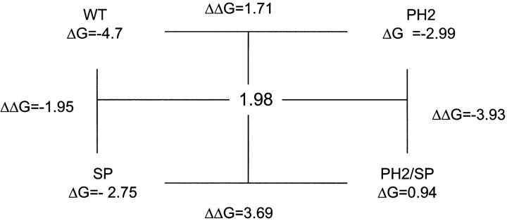
Ion channels can be gated by various extrinsic cues, such as voltage, pH, and second messengers. However, most ion channels display extrinsic cue-independent transitions as well. These events represent spontaneous conformational changes of the channel protein. The molecular basis for spontaneous gating and its relation to the mechanism by which channels undergo activation gating by extrinsic cue stimulation is not well understood. Here we show that the proximal pore helix of inwardly rectifying (Kir) channels is partially responsible for determining spontaneous gating characteristics, affecting the open state of the channel by stabilizing intraburst openings as well as the bursting state itself without affecting K(+) ion-channel interactions. The effect of the pore helix on the open state of the channel is qualitatively similar to that of two well-characterized mutations at the second transmembrane domain (TM2), which stabilize the channel in its activated state. However, the effects of the pore helix and the TM2 mutations on gating were additive and independent of each other. Moreover, in sharp contrast to the two TM2 mutations, the pore helix mutation did not affect the functionality of the agonist-responsive gate. Our results suggest that in Kir channels, the bottom of the pore helix and agonist-induced conformational transitions at the TM2 ultimately stabilize via different pathways the open conformation of the same gate.


Figures

FIGURE 1
FIGURE 1
Characterization of single-channel kinetics of Kir3.1/3.4 + Gβγ and Kir2.1 (A) Single-channel traces of Kir2.1 and Kir3.1/3.4 + Gβγ. The dotted line denotes the closed channel current level. The currents were recorded at −100 mV. Pipette solution contained 90 K with 50 μM Gd3+. (B) Open-time distribution for Kir2.1 at −100 mV. (C) Open-time distribution for Kir3.1/3.4 + Gβγ at −100 mV. For B and C, the bars are binned data events and the solid lines are curve fits. The arrows point to the peaks of the exponential fit.
FIGURE 2
FIGURE 2
Chimeric approach to study spontaneous gating in Kir channels. (A) Schematic diagram of the Kir2.1/Kir3.1 chimeras amino acid sequence. The location of the first and second transmembrane domains, M1 and M2, and the pore region, H5, are shown above the chimeras. The name of each chimera is indicated at its left, and the number of amino acid substitutions in each is indicated in parenthesis. (B) The average open time for each channel or chimera at −100 mV. (C) Open probability at −100 mV (D) Single-channel current level at −100 mV for each channel or chimera. (E) Left, a 3D model of Kir2.1 backbone based on the published structure of KcsA (Doyle et al., 1998). Q140 and T141 side chains are shown as space fill. Right, comparison between the amino acid sequences of the pore regions of Kir2.1, 3.1, and 3.4. The two amino acids composing the PH2 region are framed.
FIGURE 3
FIGURE 3
Kir2.1(PH2) displays Kir3.x-like gating. (A) Single-channel trace of Kir2.1(PH2) at −100 mV. The bottom trace is an expansion of a 500-ms segment (indicated by a horizontal bar) from the upper trace. (B) A 500-ms trace of a single Kir2.1 channel at −100 mV. (C) A 500-ms trace of a single Kir3.1/3.4 channel at −100 mV. The dotted line denotes the closed channel current level. (D) Burst-duration distribution for Kir2.1(PH2). The bars are binned events, the solid line is a curve fit. (E) Po at −100 mV for the parental Kir2.1, Kir3.1/3.4 + Gβγ, Kir2.1(PH2), Kir2.1(Q140E), and Kir2.1(T141A).
FIGURE 4
FIGURE 4
Reverse PH2 chimera shows increase in open times and burst durations. This phenotype can be attributed to E141Q\E147Q. (AC) Single-channel traces (left), open-time distributions (middle), and burst-duration distributions of bursts containing more than one opening (right) at −100 mV for (A) Kir3.1/3.4(PH2) + Gβγ, (B) Kir3.1/3.4(EQ) + Gβγ, and (C) Kir3.1/3.4 (A/T) + Gβγ. For an easy comparison, the fits of the open-time distributions (D) and burst-duration distributions (E) for the wild-type and the mutants were normalized such that the area under the plot equals 1 and superimposed.
FIGURE 5
FIGURE 5
Effect of the PH2 region on gating is additive to the effect of the S170P/S176P and C179A/C185A mutations in the TM2. All the recordings were made at −100 mV. (AB) Single-channel recordings of (A) Kir3.1/3.4(SP) and (B) Kir3.1/3.4(PH2 SP). The dotted lines mark closed channel current levels. (CD) Burst-duration distributions (of bursts containing more than one opening) for Kir3.1/3.4(SP) and Kir3.1/3.4(PH2/SP), respectively. The gray bars are binned events and the solid line is a curve fit. (E) For an easy comparison, the fits of the burst-duration distributions for the wild-type and the mutants were normalized such that the area under the plot equals 1 and superimposed. (FG) Single-channel recordings of Kir3.1/3.4(CA) and Kir3.1/3.4(PH2/CA), respectively. The dotted lines mark closed channel current levels. (HI) Burst-duration distributions (of bursts containing more than one opening) for Kir3.1/3.4(CA) and Kir3.1/3.4(PH2/CA), respectively. The gray bars are binned events and the solid line is a curve fit. (J) For an easy comparison, the fits of the burst-duration distributions for the wild-type and the CA and PH2 CA mutants were normalized such that the area under the plot equals 1 and superimposed.
FIGURE 6
FIGURE 6
PH2 region and the two TM2 mutations increase the probability of bursting and open probability in an additive manner. (A) Probability of entering a bursting state once the channel opens. (B) Open probability. The single asterisks indicate significant difference from the wild-type. Double asterisks indicate significant difference of combined PH2 and TM2 mutants over the levels of either of the respective PH2 and TM2 mutants.
Figure 10
Figure 10
Figure 11
Figure 11
FIGURE 7
FIGURE 7
Agonist activation of the channel is not affected by the pore helix mutations. Muscarinic type 2 receptor activation by 3 μM carbachol of Kir3.1/3.4 and its mutants at −80 mV. Carbachol induction levels were determined as fold increase in current over the basal current before the application of carbachol.
FIGURE 8
FIGURE 8
Potassium ion-channel interaction is not affected by the pore helix mutation E141Q/E147Q. (A) Current traces recorded at a holding potential of −80 mV from inside-out patches containing Kir3.1/3.4 channels under symmetrical K+ concentrations (in mM) 30 (top), 90 (middle), and 300 (bottom). (B) Current-to-voltage plot of single Kir3.1/3.4 (circles) and Kir3.1/3.4(EQ) (triangles) at 30 (open symbols) and 300 mM K+ (filled symbols). The solid lines are linear regressions to the data. (C) The dependence of single-channel conductance on the symmetrical K+ concentrations. The solid line is a curve fit. Data are presented as mean ± SD.
FIGURE 9
FIGURE 9
Open-time and burst duration are coupled in Kir3.1/3.4. (A) Average burst duration at −100 mM plotted as function of each mean open-time component. The solid line is a linear regression. (B) Fold increase over wild-type in τ03 and number of openings per burst for the various mutants. (C) Number of openings per burst plotted as function of τo3. The solid line is a linear regression.

References

    1. Alagem, N., M. Dvir, and E. Reuveny. 2001. Mechanism of Ba(2+) block of a mouse inwardly rectifying K+ channel: differential contribution by two discrete residues. J Physiol. 534:381–393. - PMC - PubMed
    1. Chapman, M. L., H. M. VanDongen, and A. M. VanDongen. 1997. Activation-dependent subconductance levels in the drk1 K channel suggest a subunit basis for ion permeation and gating. Biophys. J. 72:708–719. - PMC - PubMed
    1. Choe, H., L. G. Palmer, and H. Sackin. 1999. Structural determinants of gating in inward-rectifier K+ channels. Biophys. J. 76:1988–2003. - PMC - PubMed
    1. Choe, H., H. Sackin, and L. G. Palmer. 1998. Permeation and gating of an inwardly rectifying potassium channel. Evidence for a variable energy well. J. Gen. Physiol. 112:433–446. - PMC - PubMed
    1. Choe, H., H. Sackin, and L. G. Palmer. 2001. Gating properties of inward-rectifier potassium channels: effects of permeant ions. J. Membr. Biol. 184:81–89. - PubMed

Publication types

MeSH terms

Substances