

Analysis of the intrinsic bend in the M13 origin of replication by atomic force microscopy
- PMID: 12829495
- PMCID: PMC1303096
- DOI: 10.1016/S0006-3495(03)74485-3
Analysis of the intrinsic bend in the M13 origin of replication by atomic force microscopy
Abstract
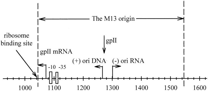
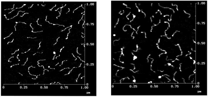
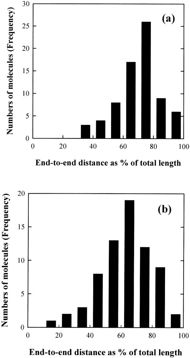
Atomic force microscopy (AFM) has been used to image a 471-bp bent DNA restriction fragment derived from the M13 origin of replication in plasmid LITMUS 28, and a 476-bp normal, unbent fragment from plasmid pUC19. The most probable angle of curvature of the 471-bp DNA fragment is 40-50 degrees, in reasonably good agreement with the bend angle determined by transient electric birefringence, 38 degrees +/- 7 degrees. The normal 476-bp DNA fragment exhibited a Gaussian distribution of bend angles centered at 0 degrees, indicating that this fragment does not contain an intrinsic bend. The persistence length, P, was estimated to be 60 +/- 8 and 62 +/- 8 nm for the 471- and 476-bp fragments, respectively, from the observed mean-square end-to-end distances in the AFM images. Since the P-values of the normal and bent fragments are close to each other, the overall flexibility of DNA fragments of this size is only marginally affected by the presence of a stable bend. The close agreement of AFM and transient electric birefringence results validates the suitability of both methods for characterizing DNA bending and flexibility.
Figures


Similar articles
-
Intrinsic curvature in the VP1 gene of SV40: comparison of solution and gel results.Biophys J. 2005 Feb;88(2):1191-206. doi: 10.1529/biophysj.104.039834. Epub 2004 Nov 19. Biophys J. 2005. PMID: 15556988 Free PMC article.
-
Analysis of DNA bending by transient electric birefringence.Biopolymers. 2003 Oct;70(2):270-88. doi: 10.1002/bip.10458. Biopolymers. 2003. PMID: 14517915
-
Intrinsic curvature of plasmid DNAs analyzed by polyacrylamide gel electrophoresis.Electrophoresis. 1996 Jun;17(6):989-95. doi: 10.1002/elps.1150170605. Electrophoresis. 1996. PMID: 8832163
-
Intrinsically bent DNA in replication origins and gene promoters.Genet Mol Res. 2008 Jun 24;7(2):549-58. doi: 10.4238/vol7-2gmr461. Genet Mol Res. 2008. PMID: 18752180 Review.
-
Is DNA a worm-like chain in Couette flow? In search of persistence length, a critical review.Sci Prog. 2009;92(Pt 2):163-204. doi: 10.3184/003685009X462205. Sci Prog. 2009. PMID: 19697713 Free PMC article. Review.
Cited by
-
Intrinsic curvature in the VP1 gene of SV40: comparison of solution and gel results.Biophys J. 2005 Feb;88(2):1191-206. doi: 10.1529/biophysj.104.039834. Epub 2004 Nov 19. Biophys J. 2005. PMID: 15556988 Free PMC article.
-
Direct measurement of DNA bending by type IIA topoisomerases: implications for non-equilibrium topology simplification.Nucleic Acids Res. 2011 Jul;39(13):5729-43. doi: 10.1093/nar/gkr109. Epub 2011 Mar 17. Nucleic Acids Res. 2011. PMID: 21421557 Free PMC article.
-
DNA sequence templates adjacent nucleosome and ORC sites at gene amplification origins in Drosophila.Nucleic Acids Res. 2015 Oct 15;43(18):8746-61. doi: 10.1093/nar/gkv766. Epub 2015 Jul 30. Nucleic Acids Res. 2015. PMID: 26227968 Free PMC article.
-
Effect of the matrix on DNA electrophoretic mobility.J Chromatogr A. 2009 Mar 6;1216(10):1917-29. doi: 10.1016/j.chroma.2008.11.090. Epub 2008 Dec 6. J Chromatogr A. 2009. PMID: 19100556 Free PMC article. Review.
-
Analysis of scanning force microscopy images of protein-induced DNA bending using simulations.Nucleic Acids Res. 2005 Apr 20;33(7):e68. doi: 10.1093/nar/gni073. Nucleic Acids Res. 2005. PMID: 15843682 Free PMC article.
References
-
- Bednar, J., P. Furrer, V. Katritch, A. Z. Stasiak, J. Dubochet, and A. Stasiak. 1995. Determination of DNA persistence length by cryo-electron microscopy. Separation of the static and dynamic contributions to the apparent persistence length of DNA. J. Mol. Biol. 254:579–594. - PubMed
-
- Bell, S. P. 2002. The origin recognition complex from simple origins to complex functions. Genes Dev. 16:659–672. - PubMed
-
- Bevington, P. R. 1969. Data Reduction and Error Analyses for the Physical Sciences. McGraw-Hill, New York.
-
- Bloomfield, V. A., D. M. Crothers, and I. Tinoco, Jr. 1999. Nucleic Acids: Structures, Properties and Functions. University Science Books, Sausalito, CA.
-
- Crothers, D. M., T. E. Haran, and J. G. Nadeau. 1990. Intrinsically bent DNA. J. Biol. Chem. 265:7093–7096. - PubMed
Publication types
MeSH terms
Grants and funding
LinkOut - more resources
Full Text Sources
Miscellaneous
