. 2003 Jul 8;100(14):8520-5.
doi: 10.1073/pnas.1332574100. Epub 2003 Jun 26.

A neuronal network model linking subjective reports and objective physiological data during conscious perception

Affiliations

A neuronal network model linking subjective reports and objective physiological data during conscious perception

Stanislas Dehaene et al. Proc Natl Acad Sci U S A. .

Abstract

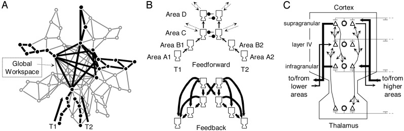
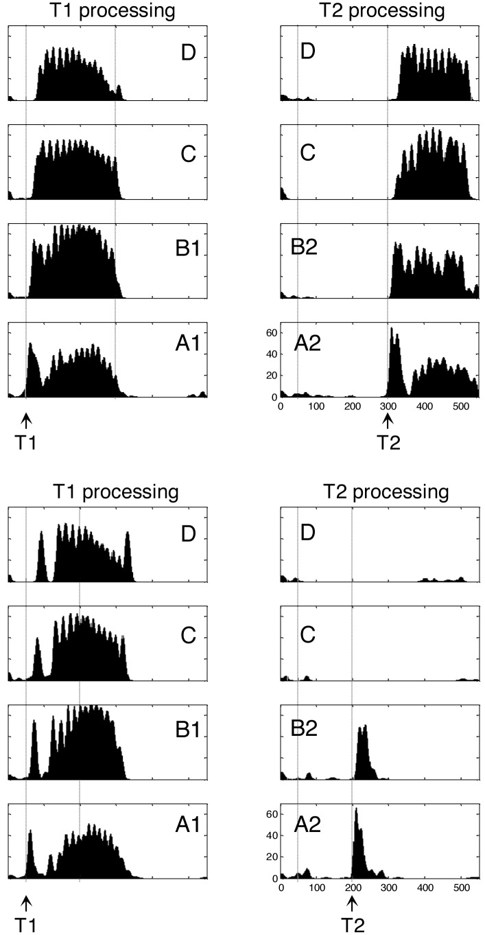
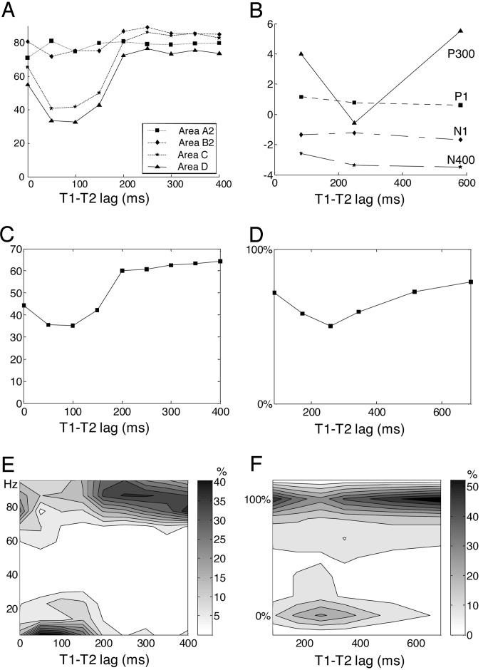
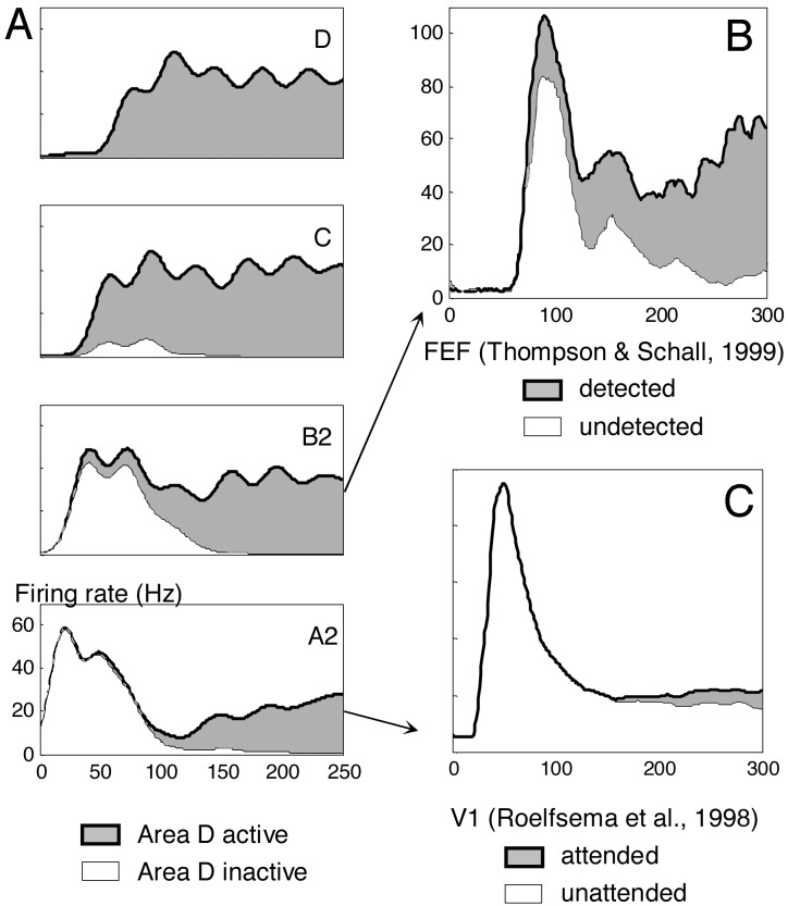
The subjective experience of perceiving visual stimuli is accompanied by objective neuronal activity patterns such as sustained activity in primary visual area (V1), amplification of perceptual processing, correlation across distant regions, joint parietal, frontal, and cingulate activation, gamma-band oscillations, and P300 waveform. We describe a neuronal network model that aims at explaining how those physiological parameters may cohere with conscious reports. The model proposes that the step of conscious perception, referred to as access awareness, is related to the entry of processed visual stimuli into a global brain state that links distant areas including the prefrontal cortex through reciprocal connections, and thus makes perceptual information reportable by multiple means. We use the model to simulate a classical psychological paradigm: the attentional blink. In addition to reproducing the main objective and subjective features of this paradigm, the model predicts an unique property of nonlinear transition from nonconscious processing to subjective perception. This all-or-none dynamics of conscious perception was verified behaviorally in human subjects.


Figures

Fig. 1.
Fig. 1.
Neuronal workspace model of conscious access. (A) Schematic architecture of brain areas (redrawn from ref. 14) comprising multiple specialized processors and a central network of high-level areas temporarily interconnecting them. During the attentional blink, T1 invades the workspace, where areas lock into a single assembly supporting conscious reportability. This invasion by T1 blocks the processing of T2 at a similar depth. (B) Subset of thalamo–cortical columns included in the present simulation. (C) Structure of a single simulated thalamo–cortical column, reproducing the laminar distribution of projections between excitatory neurons (triangles) and inhibitory neurons (stars; see Materials and Methods for details).
Fig. 2.
Fig. 2.
Simulation of two trials of the attentional blink task. In each case, boxes show the temporal evolution of firing rate in excitatory cortical neurons within each of the eight columns of Fig. 1B. (Upper) “Seen” trial with long T1–T2 lag (250 ms). T2 is presented at a time when the sustained activity evoked by T1 has decayed. T2 is therefore able to invade all cortical levels and elicits long-lasting activity comparable to T1. (Lower) “Blinked” trial with short T1–T2 lag (150 ms). When T2 is presented during T1-evoked activity, it fails to evoke neural activity beyond an initial bottom-up activation in areas A and B.
Fig. 3.
Fig. 3.
Comparison of network simulations (A, C, and E) with experimental subjective and objective data on T2 processing (B, D, and F). (A) In the simulation, the peak firing rate of T2 neurons (shown in Hz) is unaffected by T1–T2 lag in low-level areas but shows a temporary drop at intermediate lags in higher areas C and D. (B) Likewise, experimentally recorded event-related potentials evoked by T2 (shown in μV) exhibit a preservation of P1, N1, and N400 components but a drop of the P300 waveform during the attentional blink [redrawn from data by Vogel, et al. (12)]. (C) Mean power in the γ-band (20–100 Hz) emitted by simulated pyramidal neurons during a 200-ms window after T2 presentation. (E) Distribution of firing in area D. In the simulation, reverberant activation of workspace neurons is all-or-none such that parameters such as peak firing rate and γ-band power are distributed bimodally. Such a bimodal distribution was observed in an experiment in which we collected subjective ratings of T2 perception from 0% (not seen) to 100% (maximal visibility) (D, mean rating; F, distribution of ratings).
Fig. 4.
Fig. 4.
Neural activity evoked by seen and unseen T2 targets in simulation (A) and actual experimental recordings (B and C). For lags between 0 and 200 ms, simulated trials were sorted as a function of the amount of pyramidal neuron activity that reached area D in a 250-ms window after T2 presentation. (A) Averages of firing rate as a function of time show that trials with area D activity are characterized by long-lasting amplification in lower areas C, B2, and A2. (B) The curves from assembly B2, showing earlier amplification and a small difference in the amplitude of the initial peak, are similar to activity evoked in monkey frontal eye field area during a masking paradigm [redrawn from data by Thompson and Schall (34)]. (C) The curves from assembly A2, with unchanged initial peak and small subsequent amplification, are similar to actual electrophysiological recordings from monkey area V1 during attention and conscious perception paradigms [redrawn from data by Roelfsema et al. (39)].

References

    1. Super, H., Spekreijse, H. & Lamme, V. A. (2001) Nat. Neurosci. 4 304-310. - PubMed
    1. Lamme, V. A. & Roelfsema, P. R. (2000) Trends Neurosci. 23 571-579. - PubMed
    1. Lamme, V. A., Zipser, K. & Spekreijse, H. (2002) J. Cognit. Neurosci. 14 1044-1053. - PubMed
    1. Dehaene, S., Naccache, L., Cohen, L., Le Bihan, D., Mangin, J. F., Poline, J. B. & Rivière, D. (2001) Nat. Neurosci. 4 752-758. - PubMed
    1. Moutoussis, K. & Zeki, S. (2002) Proc. Natl. Acad. Sci. USA 99 9527-9532. - PMC - PubMed

Publication types