. 2003 Jul;77(14):7764-78.
doi: 10.1128/jvi.77.14.7764-7778.2003.

Mutations within the ADP (E3-11.6K) protein alter processing and localization of ADP and the kinetics of cell lysis of adenovirus-infected cells

Affiliations

Mutations within the ADP (E3-11.6K) protein alter processing and localization of ADP and the kinetics of cell lysis of adenovirus-infected cells

Ann E Tollefson et al. J Virol. 2003 Jul.

Abstract

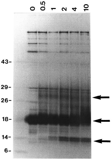
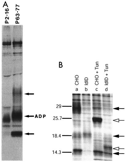
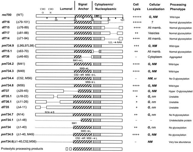
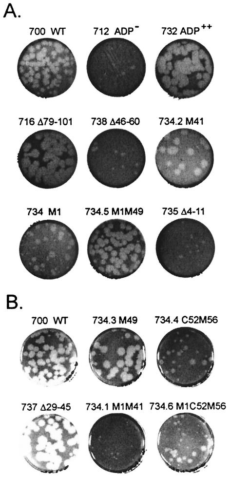
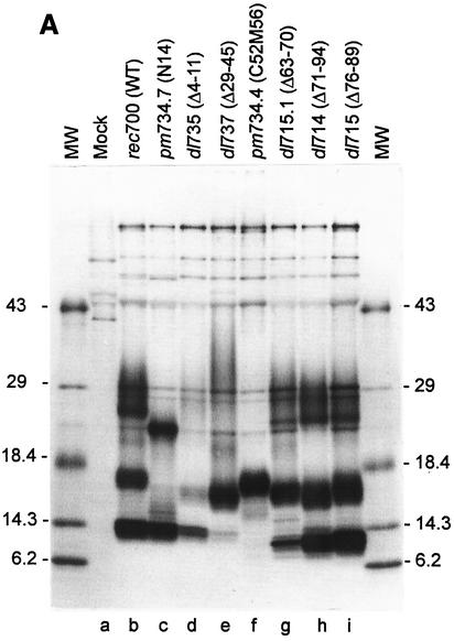
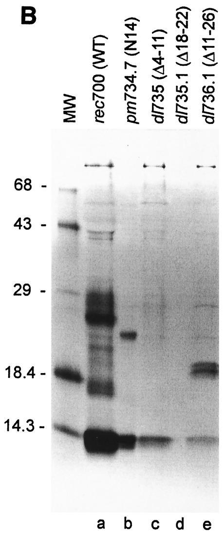
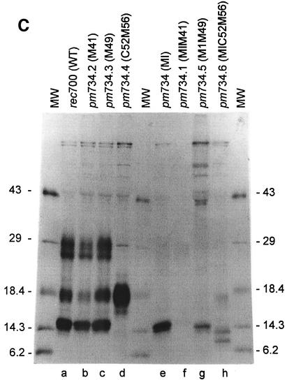
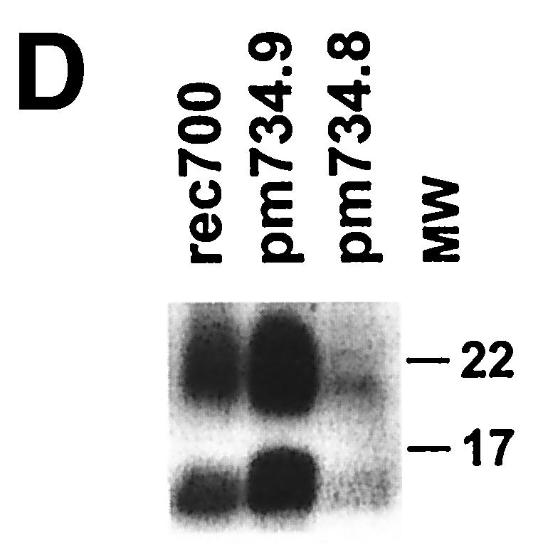
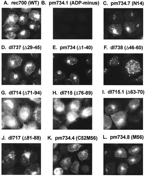
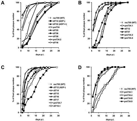
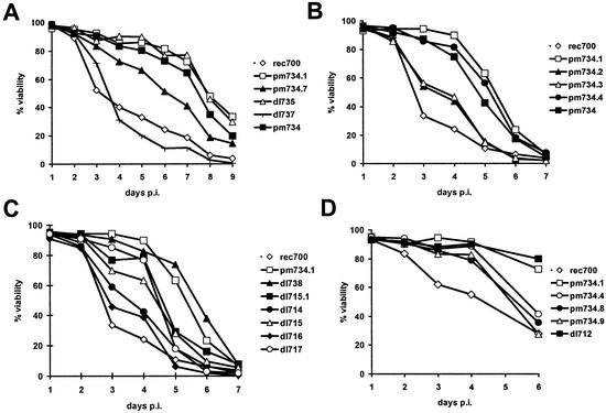
ADP (also known as E3-11.6K protein) is synthesized abundantly in late adenovirus infection and is required for efficient lysis of infected cells and release of viral progeny at the end of the viral replication cycle. ADP is a type III bitopic N(endo)C(exo) nuclear membrane and Golgi glycoprotein that is produced at high levels in late adenovirus infection (>24 h postinfection). We show pulse-chase and other studies indicating that ADP undergoes a complex process of N- and O-linked glycosylation and proteolytic cleavage. In order to further characterize ADP, a series of 23 deletion and point mutations has been constructed in the adenovirus serotype 2 adp gene and then built into a wild-type adenovirus background. These mutants were analyzed for processing and intracellular localization of ADP. Mutation of the single predicted N glycosylation site eliminated N glycosylation. Deletion of a region in ADP rich in serine and threonine residues reduced O glycosylation. In general, mutations within the lumenal domain of ADP resulted in lower protein stability; immunofluorescence assays indicated that these ADPs were primarily present in the Golgi apparatus. Viruses with mutations within the cytoplasmic-nucleoplasmic domain of ADP showed normal glycosylation patterns and protein abundance for ADP, but the protein was often found throughout cellular membranes rather than being localized specifically to the nuclear membrane and Golgi apparatus. The ADP virus mutants were analyzed by cell viability assays to determine the kinetics of cell lysis following infection of human A549 cells. In general, viruses with mutations within the lumenal domain of ADP display greatly reduced efficiencies of cell lysis. Viruses with large deletions in the cytoplasmic-nucleoplasmic domain of ADP retain much of their ability to lyse infected cells.


Figures

FIG. 1.
FIG. 1.
ADP undergoes posttranslational modification, as indicated by pulse-chase analysis of Ad2 ADP. Human KB cells in suspension culture were infected with Ad2 at 50 PFU/cell. At 26 h p.i., the cells were labeled for 20 min with Tran35S, and then at the indicated hours of chase (top), proteins were extracted. ADP was immunoprecipitated with rabbit antipeptide antiserum against residues 87 to 101 of the Ad2 sequence and separated by SDS-PAGE (10 to 18% gradient gel). The upper, middle, and lower arrows indicate the ∼27 to 31K, ∼19K, and ∼13 to 14K groups of bands of processed ADP that are discussed in the text.
FIG. 2.
FIG. 2.
Characterization of the different forms of ADP visible on SDS-PAGE. (A) Only the ∼19K form of ADP is recognized by antibody to residues 2 to 16 of the Ad2 sequence. The rec700 ADP was immunoprecipitated using rabbit antipeptide antiserum against residues 2 to 16 (lane P2-16) or 63-77 (lane P63-77) of the Ad2 ADP sequence. Lane P63-77 has forms similar to those seen in Fig. 1 (in which ADP was immunoprecipitated with antipeptide antiserum raised against residues 87 to 101 of the Ad2 sequence), while the 2-16 antibody recognized only the ∼19K form of ADP. Samples were run on 10 to 21% gradient SDS-PAGE. (B) ADP is O and N glycosylated. ADP was immunoprecipitated from Ad2-infected CHO (lanes a and c) or ldlD-14 (lanes b and d) cells. ldlD-14 cells (a CHO derivative) are deficient in O glycosylation. For lanes c and d, labeling was done in the presence of tunicamycin (+ Tun) to inhibit N glycosylation. Proteins were labeled from 24 to 30 h p.i. ADP was immunoprecipitated from cellular extracts with rabbit antiserum against residues 87 to 101 of the Ad2 sequence; samples were run on 10 to 18% gradient SDS-PAGE. The solid arrows indicate the positions of the WT ADP bands at 13 to 14K, ∼19K, and ∼27 to 31K. The open arrows indicate the bands seen in the presence of tunicamycin.
FIG. 3.
FIG. 3.
Schematic of viruses with ADP mutations. A panel of viable virus mutants with ADP deletions and point mutations was generated to evaluate the processing, localization, and function of ADP. For WT ADP (rec700), the proposed major sites for O glycosylation (five serines plus threonines) and the single N glycosylation site are shown, as are the signal-anchor and basic proline (BP) domains. The mutations present in the various viruses are indicated by the schematic. The numbers refer to the amino acid order in the ADP protein. Letters followed by a number indicate that the amino acid is mutated, e.g., L96L97L98 indicates that the Leu residues at positions 96, 97, and 98 in ADP are mutated (to Ala residues). The three columns at the right summarize the overall phenotypes of the mutants with respect to the processing of ADP, as well as the effects that the mutations have on the ability of ADP to promote cell lysis. Estimates of relative cell lysis as judged from plaque development (Fig. 7), trypan blue exclusion (Fig. 8), and lactate dehydrogenase release (not shown) data are indicated as +++++ for rec700 (WT) to − for adp-null mutants. G, Golgi apparatus; nm or NM, nuclear membrane; er or ER, endoplasmic reticulum; memb., membranes. Boldface uppercase italics indicate the primary site of localization. Lowercase letters indicate minor sites of localization.
FIG. 4.
FIG. 4.
Plaque morphology of viruses containing ADP mutations. Photographs were taken of representative dishes 14 days p.i. When plaque size for the mutants is compared to that for rec700 (the WT parental virus) and dl712 (in which adp is deleted), the plaques show a range of sizes. (A) Dishes are from the same experiment shown in Fig. 7A; (B) dishes are from the same experiment shown in Fig. 7B.
FIG. 5.
FIG. 5.
ADP synthesized by Ads with mutations in the adp gene. KB cells in suspension culture were infected with the indicated viruses, and proteins were labeled with [35S]Met and [35S]Cys. Proteins were extracted, ADP was immunoprecipitated, and the immunoprecipitates were analyzed by SDS-PAGE. The nature of the mutation for each virus is shown above each lane. (A to C) Cells were infected with 50 (A and C) or 100 (B) PFU of the mutant viruses/cell. At ∼24 h p.i., the cells were labeled with 50 (A and C) or 75 (B) μCi of [35S]Met for 4 h. In addition to the [35S]Met, 50 μCi of [35S]Cys was added for the Met point mutants (C) to compensate for the decreased number of Met residues. ADP was immunoprecipitated from cell extracts using a rabbit polyclonal antibody to residues 87 to 101 of the Ad2 ADP sequence. Mock, mock infected. (D) Immunoblot of rec700, pm734.8 (Met56 to Leu), and pm734.9 (Leu96Leu97Leu98 to AlaAlaAla). KB cells in suspension culture were infected with 50 PFU of the indicated viruses/cell and were harvested at 28.5 h p.i. For the immunoblot, a rabbit antipeptide antiserum against residues 63 to 77 of the Ad2 ADP sequence was used.
FIG. 5.
FIG. 5.
ADP synthesized by Ads with mutations in the adp gene. KB cells in suspension culture were infected with the indicated viruses, and proteins were labeled with [35S]Met and [35S]Cys. Proteins were extracted, ADP was immunoprecipitated, and the immunoprecipitates were analyzed by SDS-PAGE. The nature of the mutation for each virus is shown above each lane. (A to C) Cells were infected with 50 (A and C) or 100 (B) PFU of the mutant viruses/cell. At ∼24 h p.i., the cells were labeled with 50 (A and C) or 75 (B) μCi of [35S]Met for 4 h. In addition to the [35S]Met, 50 μCi of [35S]Cys was added for the Met point mutants (C) to compensate for the decreased number of Met residues. ADP was immunoprecipitated from cell extracts using a rabbit polyclonal antibody to residues 87 to 101 of the Ad2 ADP sequence. Mock, mock infected. (D) Immunoblot of rec700, pm734.8 (Met56 to Leu), and pm734.9 (Leu96Leu97Leu98 to AlaAlaAla). KB cells in suspension culture were infected with 50 PFU of the indicated viruses/cell and were harvested at 28.5 h p.i. For the immunoblot, a rabbit antipeptide antiserum against residues 63 to 77 of the Ad2 ADP sequence was used.
FIG. 6.
FIG. 6.
Indirect immunofluorescence assay of ADP for adp mutants. Human A549 cells growing in monolayers were infected with 50 PFU of the indicated viruses/cell. The cells were fixed in methanol at 30 h p.i. The primary antibody was rabbit antipeptide antiserum specific for residues 87 to 101 of the Ad2 sequence. The secondary antibody was goat anti-rabbit IgG (fluorescein isothiocyanate conjugate). dl717 is shown at higher magnification. The images were taken with a 60× lens, except that for dl717 for which a 100× lens was used.
FIG. 7.
FIG. 7.
The kinetics of plaque development on human A549 monolayers are altered for many Ad ADP mutants. The y axis shows the number of plaques observed on any given day of the plaque assay (x axis) as a percentage of the total number of plaques that were observed on the final day (days 25 to 30) of the plaque assay. The WT parental (rec700) and dl712 (total deletion of ADP) or pm734.1 (mutation of Met1 and Met41 in the ADP sequence; this ADP is nonfunctional) viruses are included as controls in each plaque assay. Each point represents the percentage of the final plaques that were macroscopically visible on the indicated day p.i. (A) dl732 (which overexpresses WT ADP due to altered mRNA splicing), rec700 (WT), dl712 (ΔADP), pm734 (Δ1-40), dl735 (Δ4-11), dl736 (frameshift after the first 10 codons of ADP), dl738 (Δ46-60), pm734.2 (Met41 to Leu), and dl716 (Δ79-101). (B) rec700 (WT), dl712 (ΔADP), dl737 (Δ29-45), pm734.3 (Met49 to Leu), pm734.4 (Met56 to Leu and Cys52 to Arg), and pm734.1 (Met1 and Met41 to Leu). (C) rec700 (WT), dl712 (ΔADP), dl714 (Δ71-94), dl715 (Δ76-89), dl716 (Δ79-101), dl717 (Δ81-88), pm734.7 (Asn14 to Ser), and dl715.1 (Δ63-70). (D) rec700 (WT), pm734.1 (Met1 and Met41 to Leu), pm734.4 (Met56 to Leu and Cys52 to Arg), pm734.8 (Met56 to Leu), and pm734.9 (Leu96Leu97Leu98 to Ala96Ala97Ala98).
FIG. 8.
FIG. 8.
Loss of cell viability is altered for human A549 cells infected by viruses with ADP mutations. (A) A549 cells were infected at 20 PFU per cell with the indicated viruses with ADP lumenal mutations. At daily intervals, cell viability was assayed by trypan blue exclusion. rec700 is the WT ADP control. pm734.1 is the ADP control. The virus mutations were dl735 (Δ4-11), pm734.7 (Asn14 to Ser), pm734 (Δ1-40), pm734.1 (missense mutations of Met1 and Met41 such that ADP would initiate at Met49), and dl737 (Δ29-45). (B) A549 cells were infected at 25 PFU per cell with viruses with point mutations of Met residues. The mutations were as follows: rec700 (WT control), pm734.1 (Δ1-48), pm734.2 (Met41 to Leu), pm734.3 (Met49 to Leu), pm734.4 (Met56 to Leu and Cys52 to Arg), and pm734 (Δ1-40). (C) A549 cells were infected at 25 PFU per cell with viruses containing deletions of the transmembrane or cytoplasmic sequence of ADP. The viruses were as follows: pm734.1 (Δ1-48), dl738 (Δ46-60), dl715.1 (Δ63-70), dl714 (Δ71-94), dl715 (Δ76-89), dl716 (Δ79-101), and dl717 (Δ81-88). (D) A549 cells were infected at 25 PFU per cell with viruses containing point mutations in the transmembrane or cytoplasmic sequence of ADP. Controls were rec700 (WT), pm734.1 (ADP), and dl712 (ADP). Point mutations were as follows: pm734.4 (Met56 to Leu and Cys52 to Arg), pm734.8 (Met56 to Leu), and pm734.9 (Leu96Leu97Leu98 to Ala96Ala97Ala98). Panels B and C are from the same experiment.

Similar articles

Cited by

References

    1. Bhat, B. M., H. A. Brady, and W. S. M. Wold. 1985. Virus deletion mutants that affect a 3′ splice site in the E3 transcription unit of adenovirus 2. Mol. Cell. Biol. 5:2405-2413. - PMC - PubMed
    1. Corse, E., and C. E. Machamer. 2002. The cytoplasmic tail of infectious bronchitis virus E protein directs Golgi targeting. J. Virol. 76:1273-1284. - PMC - PubMed
    1. Deutscher, S. L., B. M. Bhat, M. H. Pursley, C. Cladaras, and W. S. M. Wold. 1985. Novel deletion mutants that enhance a distant upstream 5′ splice in the E3 transcription unit of adenovirus 2. Nucleic Acids Res. 13:5771-5788. - PMC - PubMed
    1. Doronin, K., M. Kuppuswamy, K. Toth, A. E. Tollefson, P. Krajcsi, V. Krougliak, and W. S. M. Wold. 2001. Tissue-specific, tumor-selective, replication-competent adenovirus vector for cancer gene therapy. J. Virol. 75:3314-3324. - PMC - PubMed
    1. Doronin, K., K. Toth, M. Kuppuswamy, P. Krajcsi, A. E. Tollefson, and W. S. M. Wold. 2003. Overexpression of the ADP (E3-11.6K) protein increases cell lysis and spread of adenovirus. Virology 305:378-387. - PubMed

Publication types

MeSH terms

Substances

LinkOut - more resources