. 2003 Jul;77(14):8061-71.
doi: 10.1128/jvi.77.14.8061-8071.2003.

Multiple interactions across the surface of the gp120 core structure determine the global neutralization resistance phenotype of human immunodeficiency virus type 1

Affiliations

Multiple interactions across the surface of the gp120 core structure determine the global neutralization resistance phenotype of human immunodeficiency virus type 1

Peter Bouma et al. J Virol. 2003 Jul.

Abstract

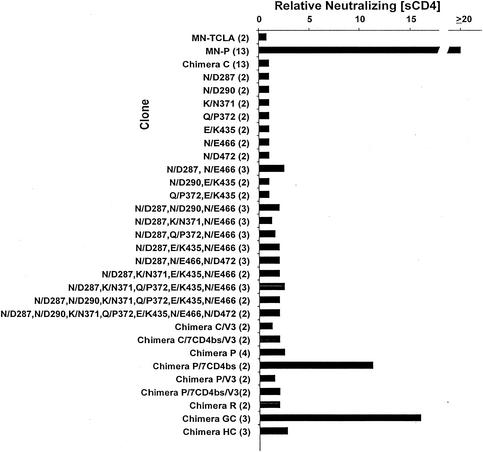
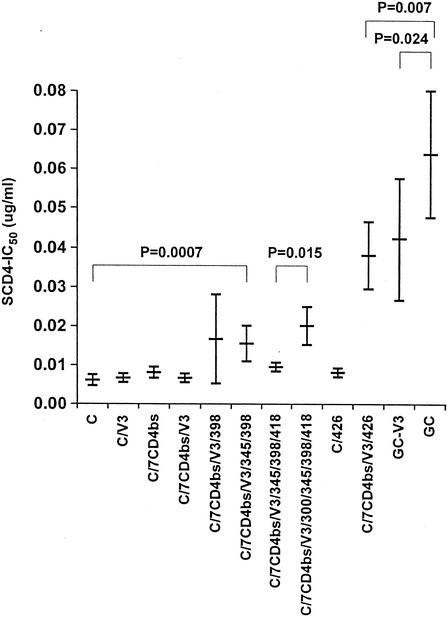
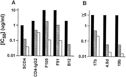
Resistance to neutralization is an important characteristic of primary isolates of human immunodeficiency virus type 1 (HIV-1) that relates to the potential for successful vaccination to prevent infection and use of immunotherapeutics for treatment of established infection. In order to further elucidate mechanisms responsible for neutralization resistance, we studied the molecular mechanisms that determine the resistance of the primary virus isolate of the strain HIV-1 MN to neutralization by soluble CD4 (sCD4). As is the case for the global neutralization resistance phenotype, sCD4 resistance depended upon sequences in the amino-terminal heptad repeat region of gp41 (HR1), as well as on multiple functional interactions within the envelope complex. The functional interactions that determined the resistance included interactions between the variable loop 1 and 2 (V1/V2) region and sequences in or near the CD4 binding site (CD4bs) and with the V3 loop. Additionally, the V3 loop region was found to interact functionally with sequences in the outer domain of gp120, distant from the CD4bs and coreceptor-binding site, as well as with a residue thought to be located centrally in the coreceptor-binding site. These and previous results provide the basis for a model by which functional signals that determine the neutralization resistance, high-infectivity phenotype depend upon interactions occurring across the surface of the gp120 core structure and involving variable loop structures and gp41. This model should be useful in efforts to define epitopes that may be important for primary virus neutralization.


Figures

FIG. 1.
FIG. 1.
Schematic representations of recombinant env genes used for analysis of the genetic basis for resistance to sCD4 neutralization. (A) Gene organization. (B) Structure of chimeric genes used in the study. Segments of each clone derived from MN-TCLA are shown as shaded bars, and segments derived from MN-P are shown as white bars. The nature of mutations introduced by site-directed mutagenesis is indicated, with locations noted by vertical lines. Construction of the clones and mutagenesis procedures are described in Materials and Methods.
FIG. 2.
FIG. 2.
Neutralization by sCD4 of viruses pseudotyped with the envelopes of MN-P (□) or chimera A (○), GC (▪), HC (⧫), or C (•). Neutralization sensitivities of MN-TCLA (data not shown) and chimera C were similar. Assays were performed in triplicate, and results shown are means obtained at each sCD4 concentration. Control luminescence was determined based on infections performed in the absence of sCD4. Essentially identical results were obtained in two similar experiments.
FIG. 3.
FIG. 3.
Alignment of deduced amino acid sequences of MN-TCLA and MN-P envelopes. Regions of the envelope proteins that are indicated schematically above the sequences include conserved regions 1, 2, 3, 4, and 5 (C1, C2, C3, C4, and C5), variable regions 1/2, 3, 4, and 5 (V1/V2, V3, V4, and V5), fusion peptide (FP), heptad repeats 1 (HR1) and 2 (HR2), transmembrane segment (TM), and cytoplasmic tail (CT). Residues identified by Kwong et al. as forming direct bonds with CD4 are marked by asterisks above the sequences (13). Alignment was produced with DNAStar.
FIG. 4.
FIG. 4.
Localization of mutations distinguishing MN-P and MN-TCLA in the atomic structure of the gp120 core. The space-filling model is based on the report of Kwong et al. (13). CD4 residues are shown in green, and 17b MAb residues are shown in yellow and aqua. Most of the gp120 residues are shown in blue, except for the residues at which mutations distinguish MN-P and MN-TCLA, which are shown in red. The approximate locations of the variable loops are shown as V1 to V5. Insets indicate the portions of the trimolecular complexes that are shown in the larger views. (A) “Front” view; (B) “back” view. Mutated residues considered to be in or close to the CD4bs were 287, 290, 371, 372, 435, 466, and 472. Inner domain residues implicated in the gp41-binding site are 91, 243, 245, and 249. Mutated residues in or near the coreceptor-binding site are 219 and 426. Mutated outer domain residues distant from binding sites are 300, 345, 398, and 418.
FIG. 5.
FIG. 5.
Mutations in and around the CD4bs contribute to sCD4 resistance in the context of MN-P V1/V2 sequences. The relative neutralizing [sCD4] was determined for each clone as follows. The 50% inhibitory concentration of sCD4 was determined by linear regression analysis in Microsoft Excel, as described in Materials and Methods. The relative neutralizing [CD4] was then calculated as test clone 50% inhibitory concentration/chimera C 50% inhibitory concentration. The 50% inhibitory concentrations for MN-P and for chimera C were determined in each experiment. The number of assays for each clone is indicated in parentheses after the clone designation. The structures of chimeras C, P, GC, and HC are illustrated in Fig. 1. Individual clones were constructed that contained one or combinations of the seven CD4bs mutations. Chimeras C/7CD4bs/V3, P/7CD4bs, and P/7CD4bs/V3 each contain the seven CD4bs mutations from MN-P (shown in Fig. 3 and 4) in the respective chimeras. Chimeras C/V3, C/7CD4bs/V3, P/V3, and P/7CD4bs/V3 each contain the four MN-P V3 region mutations, shown in Fig. 3.
FIG. 6.
FIG. 6.
Resistance of chimera GC to neutralization by sCD4 depends upon sequences in V3, the mutation at residue 426 in the coreceptor-binding site, and mutations in the outer domain distant from the CD4 and coreceptor-binding sites. The construction of chimeras C, C/V3, C/7CD4bs/V3, and GC is described in Materials and Methods and in the Fig. 5 legend. Mutations at residues 298, 345, 418, and 300, in the outer domain distant from the CD4 and coreceptor-binding sites, were introduced sequentially into chimera C/7CD4bs/V3. The V/I mutation at residue 426 was introduced into both chimera C and chimera C/7CD4bs/V3. Four mutations corresponding to the MN-TCLA V3 region sequence were introduced into chimera GC to form chimera GC-V3. Results shown are means of three assays, each done in triplicate. Statistical comparisons were done using Microsoft Excel by analysis of variance (C versus C/V3 versus C/7CD4bs/V3 versus C/7CD4bs/V3/398 versus C/7CD4bs/V3/345/398) or Student t test (C/7CD4bs/V3/345/398/418 versus C/7CD4bs/V3/300/345/398/418, C/7CD4bs/V3/426 versus GC, and BC-V3 versus GC). Results shown are the means and standard deviations of three experiments, each done in triplicate. IC50, 50% inhibitory concentration.
FIG. 7.
FIG. 7.
Functional interactions between V1/V2 and residues near the CD4bs and V3 affect global neutralization resistance. The clones MN-P (black bars), chimera P/7CD4bs (crosshatched bars), and chimera P/7CD4bs/V3 (white bars) were tested for neutralization. Results shown are averages of two assays, each done in triplicate. (A) Neutralization by the anti-CD4bs ligands sCD4, CD4-IgG2, F105, F91, and b12. (B) Neutralization by antibodies directed against the coreceptor-binding site, 17b and 4.8d, and the V3 region, 19b. In this panel, the maximum concentration of each antibody used was 2.5 μg/ml. There was no neutralization of MN-P at any concentration, so the inhibitory concentration was considered to be ≥5 μg/ml. IC50, 50% inhibitory concentration.
FIG. 8.
FIG. 8.
Schematic representation of the functional interactions of the V1/V2 and V3 regions and gp41 over the surface of gp120. The view is the same as the “front” view in Fig. 4A. The cysteine residues at the stalks of the V1/V2 and V3 loops are shown in magenta, and red arrows highlight their locations. See the text for an explanation.

References

    1. Back, N. K. T., L. Smit, M. Schutten, P. L. Nara, M. Tersmette, and J. Goudsmit. 1993. Mutations in human immunodeficiency virus type 1 gp41 affect sensitivity to neutralization by gp120 antibodies. J. Virol. 67:6897-6902. - PMC - PubMed
    1. Bagley, J., P. J. Dillon, C. Rosen, J. Robinson, J. Sodroski, and W. A. Marasco. 1994. Structural characterization of broadly neutralizing human monoclonal antibodies against the CD4 binding site of HIV-1 gp120. Mol. Immunol. 31:1149-1160. - PubMed
    1. Barbas, C. F., J. E. Crowe, Jr., D. Cababa, T. M. Jones, S. L. Zebedee, B. R. Murphy, R. M. Chanock, and D. R. Burton. 1992. Human monoclonal Fab fragments derived from a combinatorial library bind to respiratory syncytial virus F glycoprotein and neutralize infectivity. Proc. Natl. Acad. Sci. USA 89:10164-10168. - PMC - PubMed
    1. Berman, P. W., T. J. Matthews, L. Riddle, M. Champe, M. R. Hobbs, G. R. Nakamura, J. Mercer, D. J. Eastman, C. Lucas, A. J. Langlois, F. M. Wurm, and T. J. Gregory. 1992. Neutralization of multiple laboratory and clinical isolates of human immunodeficiency virus type 1 (HIV-1) by antisera raised against gp120 from the MN isolate of HIV-1. J. Virol. 66:4464-4469. - PMC - PubMed
    1. Boots, L. J., P. M. McKenna, B. A. Arnold, P. M. Keller, M. K. Gorny, S. Zolla-Pazner, J. E. Robinson, and A. J. Conley. 1997. Anti-human immunodeficiency virus type 1 human monoclonal antibodies that bind discontinuous epitopes in the viral glycoproteins can identify mimotopes from recombinant phage peptide display libraries. AIDS Res. Hum. Retrovir. 13:1549-1559. - PubMed

Publication types

MeSH terms