. 2003 Jul;77(14):8108-15.
doi: 10.1128/jvi.77.14.8108-8115.2003.

Human herpesvirus 7 open reading frame U12 encodes a functional beta-chemokine receptor

Affiliations

Human herpesvirus 7 open reading frame U12 encodes a functional beta-chemokine receptor

Kazushi Nakano et al. J Virol. 2003 Jul.

Abstract

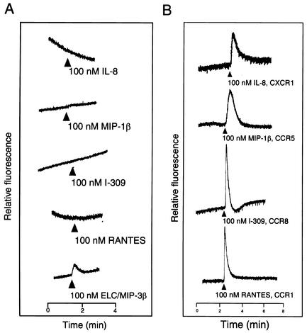
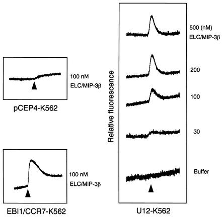
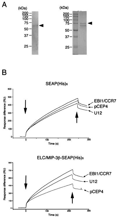
Human herpesvirus 7 (HHV-7), which belongs to the betaherpesvirus subfamily, infects mainly CD4+ T cells in vitro and infects children during infancy. After the primary infection, HHV-7 becomes latent. HHV-7 contains two genes (U12 and U51) that encode putative homologs of cellular G-protein-coupled receptors. To analyze the biological function of the U12 gene, we cloned the gene and expressed the U12 protein in cells. The U12 gene encoded a calcium-mobilizing receptor for the EBI1 ligand chemokine-macrophage inflammatory protein 3beta (ELC/MIP-3beta) but not for other chemokines, suggesting that the chemokine selectivity of the U12 gene product is distinct from that of the known mammalian chemokine receptors. These studies revealed that U12 activates distinct transmembrane signaling pathways that may mediate biological functions by binding with a beta-chemokine, ELC/MIP-3beta.


Figures

FIG. 1.
FIG. 1.
(A) Agarose gel (ethidium bromide stained) showing the results of PCR and RT-PCR amplification of HHV-7 DNA and mRNA, obtained from HHV-7-infected cell lines at day 7 postinfection. A 30-cycle PCR amplification was performed on a 1,102-bp fragment from the U12 gene of HHV-7. Lane 1, DNA size markers (φX174/HaeIII digest); lane 2, DNA from infected SupT1 cells; lane 3, cDNA from infected SupT1 cells treated with RT; lane 4, same as lane 3 but without RT. Arrowheads indicate the position of U12 and the splice variant. (B) Northern blot analysis of U12 expression in SupT1 cells infected with HHV-7. Total RNA was isolated from mock-infected and HHV-7-infected SupT1 cells on day 3 after infection. The lower panel shows ethidium bromide staining. The arrowhead indicates the U12 transcript.
FIG. 2.
FIG. 2.
Genomic structure of the U12 gene. (A) Schematic description of the U12 cDNA structure. (B) Nucleotide sequences of the exon-intron boundaries. Uppercase indicates exons; lowercase represents introns.
FIG. 3.
FIG. 3.
Sequence alignment of the U12 cDNA product and human EBI1/CCR7. Dashes indicate gaps that were inserted to optimize the alignment. Overlines mark the amino acid sequences of the transmembrane regions (I to VII). Dark and light gray boxes, identical and homologous amino acids, respectively.
FIG. 4.
FIG. 4.
Optical sectioning. (A) Confocal laser microscopy analysis and deconvolution software confirmed that K562 cells were transfected with either the pEGFP-N1 vector alone (a and b) or the U12-pEGFP fusion vector (c and d). EGFP products resulted in a predominantly cytoplasmic distribution with a strong signal, but not all cells were labeled. In contrast, the U12-GFP fusion protein accumulated in large granules that were clearly distinct from the more numerous speckles seen on the cell surface. (B) Cells were observed 24 h after transfection in two planes.
FIG. 5.
FIG. 5.
Ca2+ mobilization analysis in K562 cells transfected with U12 and treated with ELC/MIP-3β, RANTES, I-309, MIP-1β, or IL-8 (A) and K562 cells transfected with CCR1, CCR8, CCR5, or CXCR1 and treated with RANTES, I-309, MIP-1β, or IL-8 (B). Indo-1 AM-loaded cells were suspended at 2.5 × 10⁶ to 3 × 10⁶/ml and continuously monitored for fluorescence changes. Peptide agonists were added at the indicated times at a dilution of 1:25 (vol/vol), resulting in final concentrations of 100 nM. The tracings are from a single experiment that was representative of two separate experiments.
FIG. 6.
FIG. 6.
Dose-response experiments. K562 cells expressing U12 were loaded with indo-1 AM. (Right) Magnitudes of the peak transient calcium increase elicited by the indicated concentrations of MIP-3β. (Left) Parental K562 cells transfected with vector alone did not respond to 100 nM ELC/MIP-3β, while K562 cells transfected with EBI1/CCR7 did respond to 100 nM ELC/MIP-3β.
FIG. 7.
FIG. 7.
Expression of ELC/MIP-3β-SEAP-His6 and SEAP-His6 fusion proteins. (A) ELC/MIP-3β-SEAP-His6 and SEAP-His6 were purified from the conditioned medium of transfected cells by metal affinity chromatography, fractionated on 10% polyacrylamide gels, and stained with Coomassie brilliant blue. Arrowheads indicate ELC/MIP-3β-SEAP-His6 (left) and SEAP-His6 (right). The positions of size markers are shown on the left. (B) Real-time analysis of the interaction between ELC/MIP-3β or SEAP-His6 and U12-, EBI1/CCR7-, or pCEP4-expressing K562 cells, as monitored by SPR (Biacore). The SPR signal was expressed as resonance units (RU). ELC/MIP-3β-SEAP-His6 was immobilized on an NTA sensor chip, and K562 cells stably expressing U12, EBI1/CCR7, or pCEP4 were injected. Association and dissociation starts are shown by the left and right arrows, respectively. Representative binding curves for the interaction of ELC/MIP-3β with K562 cells transfected with U12 or EBI1/CCR7 and with parental pCEP4 cells are shown. The tracings are from a single experiment that was representative of three separate experiments.

References

    1. Baba, M., T. Imai, M. Nishimura, M. Kakizaki, S. Takagi, K. Hieshima, H. Nomiyama, and O. Yoshie. 1997. Identification of CCR6, the specific receptor for a novel lymphocyte-directed CC chemokine LARC. J. Biol. Chem. 272:14893-14898. - PubMed
    1. Bais, C., B. Santomasso, O. Coso, L. Arvanitakis, E. G. Raaka, J. S. Gutkind, A. S. Asch, E. Cesarman, M. C. Gershengorn, E. A. Mesri, and M. C. Gerhengorn. 1998. G-protein-coupled receptor of Kaposi's sarcoma-associated herpesvirus is a viral oncogene and angiogenesis activator. Nature 391:86-89. - PubMed
    1. Berneman, Z. N., D. V. Ablashi, G. Li, M. Eger-Fletcher, M. S. Reitz, Jr., C. L. Hung, I. Brus, A. L. Komaroff, and R. C. Gallo. 1992. Human herpesvirus 7 is a T-lymphotropic virus and is related to, but significantly different from, human herpesvirus 6 and human cytomegalovirus. Proc. Natl. Acad. Sci. USA 89:10552-10556. - PMC - PubMed
    1. Birkenbach, M., K. Josefsen, R. Yalamanchili, G. Lenoir, and E. Kieff. 1993. Epstein-Barr virus-induced genes: first lymphocyte-specific G protein-coupled peptide receptors. J. Virol. 67:2209-2220. - PMC - PubMed
    1. Black, J. B., T. F. Schwarz, J. L. Patton, K. Kite-Powell, P. E. Pellett, S. Wiersbitzky, R. Bruns, C. Muller, G. Jager, and J. A. Stewart. 1996. Evaluation of immunoassays for detection of antibodies to human herpesvirus 7. Clin. Diagn. Lab. Immunol. 3:79-83. - PMC - PubMed

Publication types