

An indirect ELISA for detection of Encephalitozoon cuniculi infection in farmed blue foxes (Alopex lagopus)
- PMID: 12831174
- PMCID: PMC1764197
- DOI: 10.1186/1751-0147-43-211
An indirect ELISA for detection of Encephalitozoon cuniculi infection in farmed blue foxes (Alopex lagopus)
Abstract
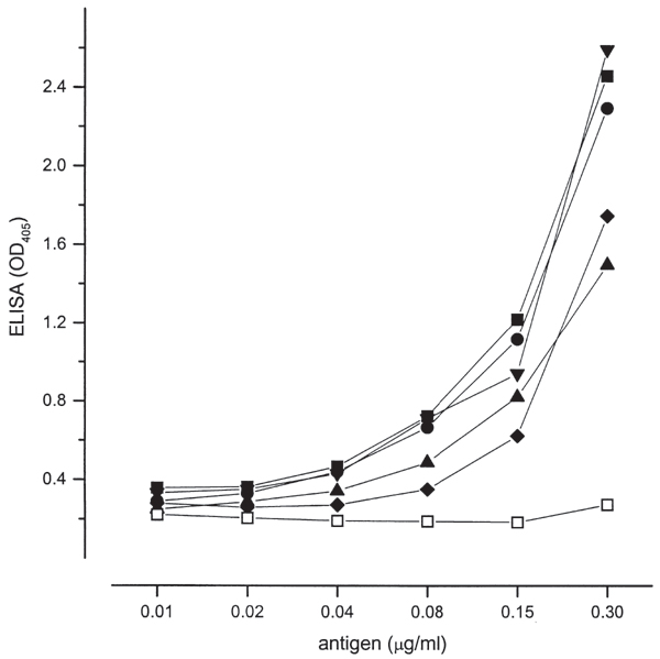
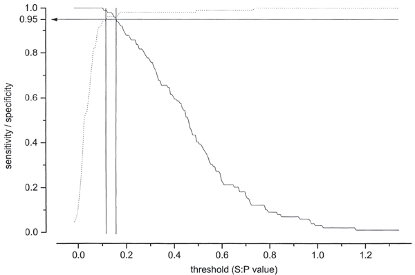
Infection with the intracellular microsporidium Encephalitozoon cuniculi can cause serious disease, encephalitozoonosis, in the blue fox (Alopex lagopus). The disease diagnosis is based on clinical signs and pathological findings, and detection of E. cuniculi or circulating antibodies directed against the parasite. Indirect immunofluorescence (IFAT) and carbon immunoassay (CIA) are the most commonly used serological methods for diagnosis in this species. In the present study, an indirect ELISA (enzyme linked immunosorbent assay) was established and evaluated against IFAT by testing of 205 field samples from blue foxes. There was high agreement between the results of the ELISA and CIA (kappa=0.99), and the ELISA and IFAT (kappa=0.958). There was no significant statistical difference between the tests (p>0.05). It was concluded that the ELISA could be used to identify seropositive farmed blue foxes. The advantage of the ELISA lies in the potential of screening large numbers of animals with the goal of eradicating E. cuniculi infection in the farms.
Figures
References
-
- Altman DG. Practical statistics for medical research. 1. Chapman & Hall, London, England; 1993. p. 611.
-
- Beckwith C, Peterson N, Liu JJ, Shadduck JA. Dot enzyme-linked immunosorbent assay (dot ELISA) for antibodies to Encephalitozoon cuniculi. Lab Anim Sci. 1988;38:573–576. - PubMed
-
- Bywater JEC, Kellett BS. The eradication of Encephalitozoon cuniculi from a specific pathogenfree rabbit colony. Lab Anim Sci. 1978;28:402–404. - PubMed
-
- Canning EU, Lom J. The microsporidia of vertebrates. Academic Press, London; 1986. p. 289.
-
- Cox JC, Gallichio HA. An evaluation of indirect immunofluorescence in the serological diagnosis of Nosema cuniculi infection. Res Vet Sci. 1977;22:50–52. - PubMed
Publication types
MeSH terms
Substances
LinkOut - more resources
Full Text Sources
