. 2003 Jul 15;17(14):1703-8.
doi: 10.1101/gad.1104803. Epub 2003 Jun 27.

IRF-4,8 orchestrate the pre-B-to-B transition in lymphocyte development

Affiliations

IRF-4,8 orchestrate the pre-B-to-B transition in lymphocyte development

Runqing Lu et al. Genes Dev. .

Abstract

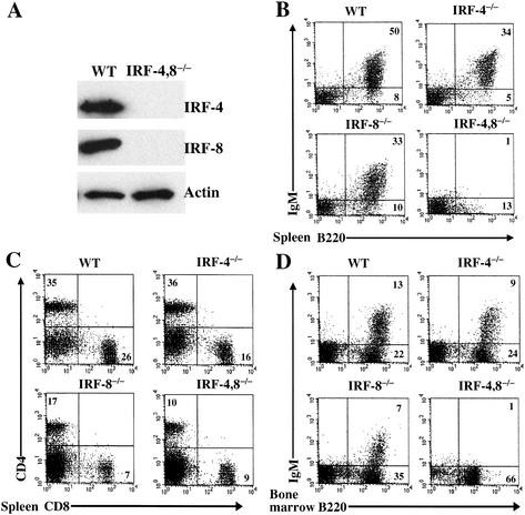
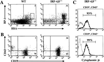
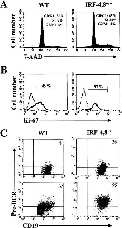
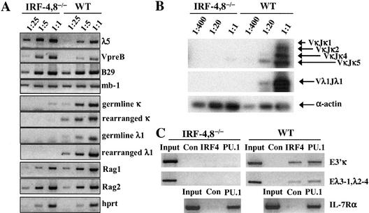
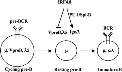
B-lymphocyte development involves sequential DNA rearrangements of immunoglobulin (Ig) heavy (mu) and light (kappa, lambda) chain loci and is dependent on transient expression of mu containing pre-antigen receptor complexes (pre-BCR). To date, genetic analysis has not identified transcription factors that coordinate the pre-B-to-B transition. We demonstrate that the related interferon regulatory factors IRF-4 (Pip) and IRF-8 (ICSBP) are required for Ig light but not heavy-chain gene rearrangement. In the absence of these transcription factors, B-cell development is arrested at the cycling pre-B-cell stage and the mutant cells fail to down-regulate the pre-BCR. On the basis of molecular analysis, we propose that IRF-4,8 function as a genetic switch to down-regulate surrogate light-chain gene expression and induce conventional light-chain gene transcription and rearrangement.


Figures

Figure 1.
Figure 1.
B-cell development is blocked in IRF-4,8-/- mice. (A) Western blot analysis of IRF-4 and IRF-8 expression in splenocytes from wild-type and IRF-4,8-/- mice. (B–D) FACS analysis of B- and T-lymphocyte populations in spleen and bone marrow of wild-type and IRF-4,8 mutant mice. Cells were examined using a lymphocyte gate and antibodies directed against B220, IgM, CD4, and CD8. Numbers in quadrants indicate percentage of cells in the gated population.
Figure 2.
Figure 2.
B-cell development is arrested at the pre-B stage in IRF-4,8-/- mice. (A) Bone marrow cells were analyzed for expression of B220, CD43, BP-1, and HSA using four-color FACS. The BP-1 and HSA profiles were obtained after gating on B220+, CD43+ cells. (B) Bone marrow cells were analyzed for CD19 and CD43 expression after gating on B220+ lymphocytes. (C) Intracellular μ expression in CD19+ CD43+, or CD19+ CD43- IRF-4,8-/- mutant B-lineage cells. Myeloid-lineage-depleted cells (Mac-1-, Gr-1-) from IRF-4,8-/- bone marrow were stained with anti-CD19 and anti-CD43 antibodies. After fixation and permeabilization, the cells were reacted with an anti-IgM antibody (solid line) or an isotype control (broken line), and then analyzed by FACS.
Figure 3.
Figure 3.
IRF-4,8-/- pre-B cells have an increased proliferation index and express higher levels of the pre-BCR. (A,B) Cell cycle status of IRF-4,8-/- pre-B cells. Bone marrow cells after enriching for sIgM—B-lineage cells were stained with anti-CD19 and anti-CD43 antibodies. After fixation and permeabilization, the cells were incubated with either 7-AAD or anti-Ki-67 antibody. FACS analysis was performed by gating on CD19+ CD43- cells. Ki-67 staining was assessed using an isotype-matched antibody as control (broken line). (C) Expression of pre-BCR on IRF-4,8-/- pre-B cells. (Top) Freshly isolated bone marrow cells were analyzed by four-color FACS using anti-CD19, CD43, IgM, and pre-BCR antibodies. The FACS profiles were obtained by gating on CD19+, CD43- and IgM-, and cells. (Bottom) The same FACS analysis was performed using pre-B cells that were expanded in culture using IL-7. For each panel, the pre-BCR quadrant was set using an isotype control.
Figure 4.
Figure 4.
IRF-4,8-/- pre-B cells express high levels of surrogate light-chain genes (λ5, VpreB) and are impaired for conventional light-chain (κ, λ) transcription and rearrangement. (A) Reverse transcriptase PCR (RT—PCR) analysis of B-lineage genes expressed in IRF-4,8-/- pre-B cells. CD19+, CD43-, and IgM- pre-B cells were isolated by sorting after enrichment of B-lineage cells from bone marrow. Serial dilutions of total cDNA were used for RT—PCR with primers for the indicated genes. (B) PCR analysis of κ and λ DNA rearrangements. Serial dilutions of genomic DNA from the cells described in A were analyzed using primers that detect the indicated κ and λ rearrangements. (C) Chromatin cross-linking and immunoprecipitation analysis of IRF-4 and PU.1 binding to the κ 3′ and λ enhancers. IL-7 expanded pre-B cells, as described in Figure 3C, were cross-linked with formaldehyde, and then chromatin fragments were immunoprecipitated using anti-IRF-4 or anti-PU.1 antibodies. Rabbit IgG antibodies were used as a control. Indicated DNA fragments were amplified by PCR. Input represents an equivalent amount of cross-linked chromatin not subjected to immunoprecipitation.
Figure 5.
Figure 5.
A model for transcriptional control of the pre-B-to-B transition. IRF-4,8 are depicted as components of a genetic switch whose function is to down-regulate expression of the surrogate light-chain genes, thereby inhibiting pre-BCR-dependent proliferation of pre-B cells. Concomitantly, IRF-4,8 promote pre-B-cell differentiation by inducing conventional light-chain gene transcription and rearrangement. These latter processes are proposed to depend on binding of IRF-4/8 to κ and λ enhancers with the transcription factors PU.1/Spi-B.

References

    1. Brass A.L., Zhu, A.Q., and Singh, H. 1999. Assembly requirements of PU.1–Pip (IRF-4) activator complexes: Inhibiting function in vivo using fused dimers. EMBO J. 18: 977–991. - PMC - PubMed
    1. Burrows P.D., Stephan, R.P., Wang, Y.H., Lassoued, K., Zhang, Z., and Cooper, M.D. 2002. The transient expression of pre-B cell receptors governs B cell development. Semin. Immunol. 14: 343–349. - PubMed
    1. Cheng A.M., Rowley, B., Pao, W., Hayday, A., Bolen, J.B., and Pawson, T. 1995. Syk tyrosine kinase required for mouse viability and B-cell development. Nature 378: 303–306. - PubMed
    1. DeKoter R.P., Lee, H.J., and Singh, H. 2002. PU.1 regulates expression of the interleukin-7 receptor in lymphoid progenitors. Immunity 16: 297–309. - PubMed
    1. Desiderio S., Lin, W.C., and Li, Z. 1996. The cell cycle and V(D)J recombination. Curr. Top. Microbiol. Immunol. 217: 45–59. - PubMed

Publication types

LinkOut - more resources