. 2003 Jul 15;17(14):1714-26.
doi: 10.1101/gad.264303. Epub 2003 Jun 27.

A novel class of secreted hydrophobic proteins is involved in aerial hyphae formation in Streptomyces coelicolor by forming amyloid-like fibrils

Affiliations

A novel class of secreted hydrophobic proteins is involved in aerial hyphae formation in Streptomyces coelicolor by forming amyloid-like fibrils

Dennis Claessen et al. Genes Dev. .

Abstract

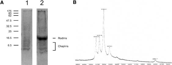
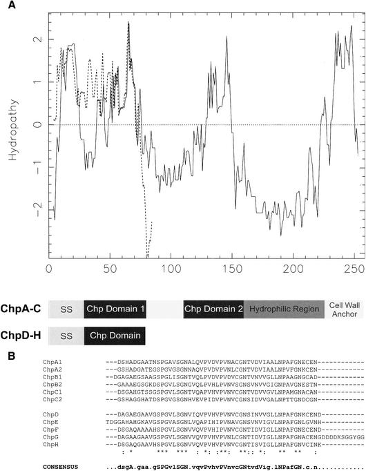
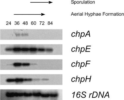
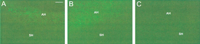
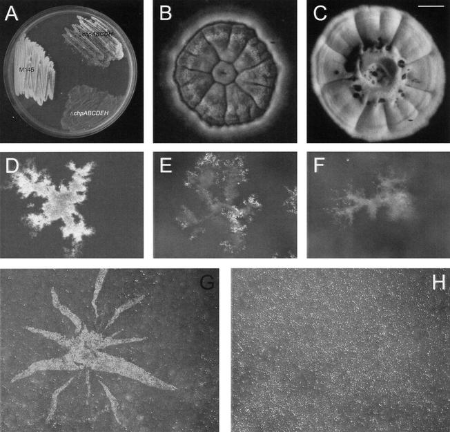
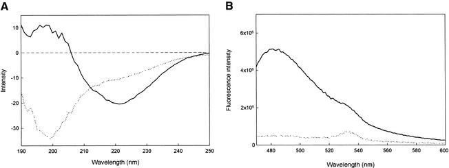
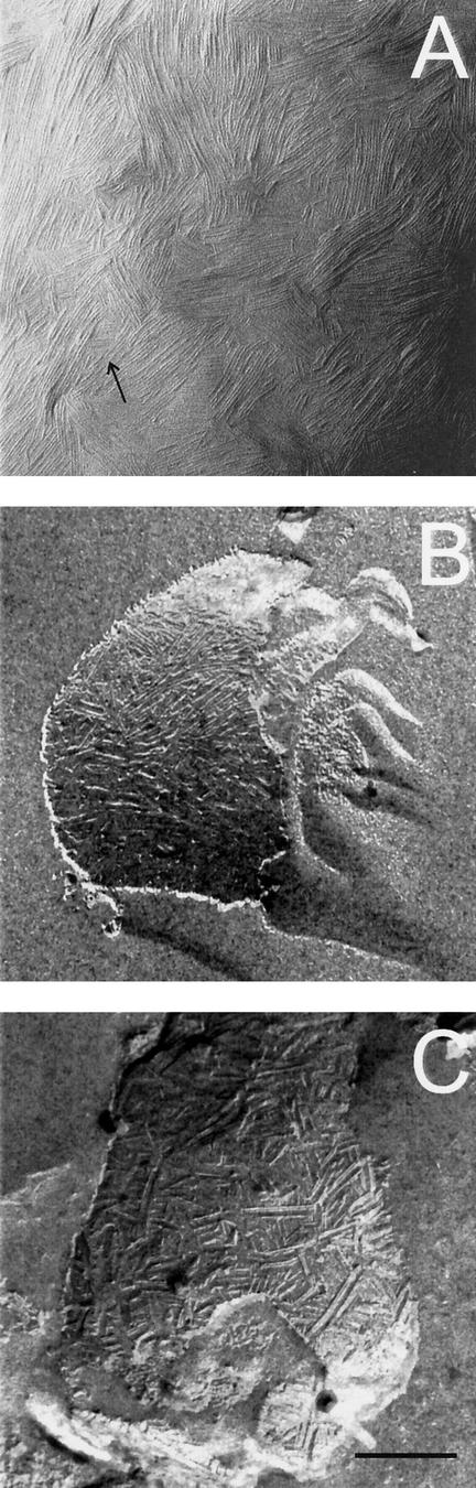
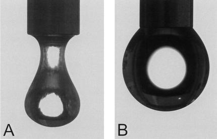
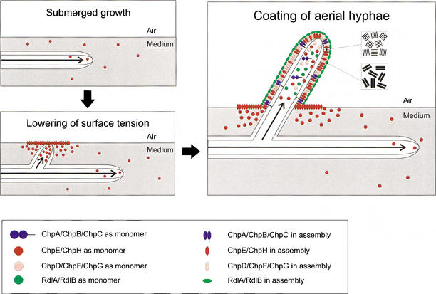
Streptomycetes exhibit a complex morphological differentiation. After a submerged mycelium has been formed, filaments grow into the air to septate into spores. A class of eight hydrophobic secreted proteins, ChpA-H, was shown to be instrumental in the development of Streptomyces coelicolor. Mature forms of ChpD-H are up to 63 amino acids in length, and those of ChpA-C are larger (+/-225 amino acids). ChpA-C contain two domains similar to ChpD-H, as well as a cell-wall sorting signal. The chp genes were expressed in submerged mycelium (chpE and chpH) as well as in aerial hyphae (chpA-H). Formation of aerial hyphae was strongly affected in a strain in which six chp genes were deleted (DeltachpABCDEH). A mixture of ChpD-H purified from cell walls of aerial hyphae complemented the DeltachpABCDEH strain extracellularly, and it accelerated development in the wild-type strain. The protein mixture was highly surface active, and it self-assembled into amyloid-like fibrils at the water-air interface. The fibrils resembled those of a surface layer of aerial hyphae. We thus conclude that the amyloid-like fibrils of ChpD-H lower the water surface tension to allow aerial growth and cover aerial structures, rendering them hydrophobic. ChpA-C possibly bind ChpD-H to the cell wall.


Figures

Figure 1.
Figure 1.
(A) SDS-PAGE analysis of TFA extracts of SDS-treated cell walls of sporulating cultures of S. coelicolor M145ΔrdlAB (lane 1) and wild-type strain M145 (lane 2). (B) A faint smear is observed between 5 and 10 kD, consisting of a mixture of five small proteins, called ChpD–H, with masses ranging from 5071 to 5994 Da as determined by MALDI-TOF mass spectrometry.
Figure 2.
Figure 2.
(A) The hydropathy patterns of ChpC (solid line) and ChpG (broken line) illustrate the hydrophobic nature of the large (ChpA–C) and small (ChpD–H) chaplins. All chaplins have a signal sequence for secretion (SS). In ChpA–C, the two regions that conform to the sequences of the small chaplins (dark boxes) are followed by a hydrophilic region and a putative cell wall anchor containing an LAXTG motif and a stretch of hydrophobic amino acids. (B) Alignment of the homologous sequences present in the eight chaplins. The consensus sequence shows residues that are present in >50% of the sequences. The conserved amino acids, present in all sequences, are indicated in uppercase.
Figure 3.
Figure 3.
Temporal expression of the chp genes in S. coelicolor grown on solid NMMP medium as determined by Northern analysis. Genes chpE and chpH were expressed after 24 h of growth, when cultures still grew submerged, and during formation of aerial hyphae. In contrast, chpA and chpF were expressed during formation of aerial hyphae only. Expression of chpA was 10- to 25-fold lower compared with that of chpE, chpF, and chpH. Blots were rehybridized with 16S rDNA to confirm that lanes contained equal amounts of RNA.
Figure 4.
Figure 4.
Spatial expression of chpC (A) and chpH (B) in S. coelicolor grown on solid medium for 2 d and visualized using eGFP as a reporter. GFP expression was observed in aerial hyphae (AH) of transformants but not in submerged hyphae (SH). Fluorescence due to the chpC promoter was relatively low compared with that of chpH. (C) Wild-type hyphae served as a control. Bar, 10 μm.
Figure 5.
Figure 5.
Chaplins are involved in formation of aerial hyphae. (A) After 3 d of growth on solid minimal medium, the wild-type strain formed a confluent layer of white aerial hyphae, whereas the ΔchpABCDH and the ΔchpABCDEH were strongly affected in aerial growth. (B) The ΔchpABCDH strain formed only few aerial hyphae on solid complete R5 medium even after 8 d of growth, the wild-type strain serving as a control (C). Colonies of the wild-type strain at the water–air interface of liquid standing minimal medium showed a confluent layer of aerial hyphae after 4 d of growth (D), in contrast to the ΔchpABCDH strain (E) and the ΔchpABCDEH strain (F). Formation of aerial hyphae, as indicated by a white fluffy layer, could be restored by applying a mixture of chaplins extracted from a 3-day-old culture of the ΔrdlAB strain at the surface of a colony of the ΔchpABCDEH strain grown on solid medium (G), an extract of the ΔchpABCDEH strain serving as a control (H). Bars: B,C,G,H, 1 mm; D–F, 400 μm.
Figure 6.
Figure 6.
Conformational changes of a mixture of chaplins (ChpD–H) isolated from cell walls of the M145ΔrdlAB strain. (A) The circular dichroism spectrum of an aqueous solution of the mixture of small chaplins was indicative for random coiled proteins (broken line), and the protein mixture adopted a conformation rich in β-sheet on vortexing (solid line). (B) Chaplins in the β-sheet rich state interact with the fluorescent dye thioflavin T (solid line), increasing its fluorescence 10-fold compared with water that served as a control (broken line).
Figure 7.
Figure 7.
(A) Four- to six-nanometer-wide fibrils are formed after drying down an aqueous solution of chaplins. The fibrils resemble those present at the outer surface of the M145ΔrdlAB strain (B) and are different from the rodlets present on aerial hyphae and spores of the wild-type strain (C). Arrow indicates direction of shadowing. Bar, 200 μm.
Figure 8.
Figure 8.
Surface activity of a mixture of chaplins (A), as indicated by the shape of an 8-μL droplet, water serving as a control (B).
Figure 9.
Figure 9.
The role of chaplins and rodlins in the formation of aerial hyphae in S. coelicolor. Initially, hyphae grow submerged, secreting ChpE and ChpH. These proteins lower the water surface tension dramatically by assembling at the water–air interface. This enables hyphae to grow into the air. Emerging aerial hyphae produce ChpA–H that assemble at the cell wall–air interface into an insoluble protein film characterized by an ultrastructure of 4–6-nm-wide fibrils. The chaplin layer provides a hydrophobic surface and this may induce the formation of the typical rodlet layer formed by RdlA and RdlB.

References

    1. Bentley S.D., Chater, K.F., Cerdeño-Tárraga, A.-M., Challis, G.L., Thomson, N.R., James, K.D., Harris, D.E., Quail, M.A., Kieser, H., Harper, D., et al. 2002. Complete genome sequence of the model actinomycete Streptomyces coelicolor A3(2). Nature 417: 141–147. - PubMed
    1. Butko P., Buford, J.P., Goodwin, J.S., Stroud, P.A., McCormick, C.L., and Cannon, G.C. 2001. Spectroscopic evidence for amyloid-like interfacial self-assembly of hydrophobin Sc3. Biochem. Biophys. Res. Commun. 280: 212–215. - PubMed
    1. Champness W.C. 1988. New loci required for Streptomyces coelicolor morphological and physiological differentiation. J. Bacteriol. 170: 1168–1174. - PMC - PubMed
    1. Chapman M.R., Robinson, L.S., Pinkner, J.S., Roth, R., Heuser, J., Hammar, M., Normark, S., and Hultgren, S.J. 2002. Role of Escherichia coli curli operons in directing amyloid fiber formation. Science 295: 851–855. - PMC - PubMed
    1. Chater K.F. 1998. Taking a genetic scalpel to the Streptomyces colony. Microbiology 144: 1465–1478. - PubMed

MeSH terms