. 2003 Jul;23(14):4841-58.
doi: 10.1128/MCB.23.14.4841-4858.2003.

C/EBPbeta regulation in lipopolysaccharide-stimulated macrophages

Affiliations

C/EBPbeta regulation in lipopolysaccharide-stimulated macrophages

Michelle N Bradley et al. Mol Cell Biol. 2003 Jul.

Abstract

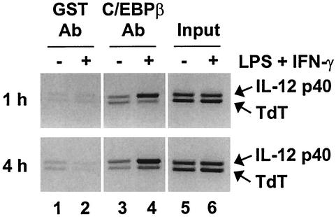
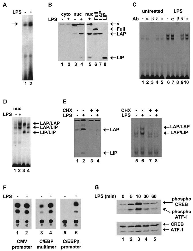
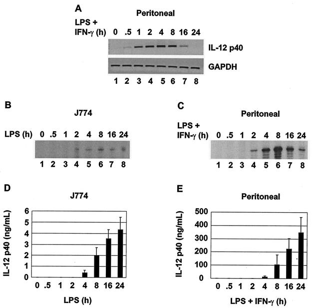
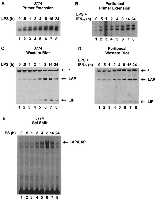
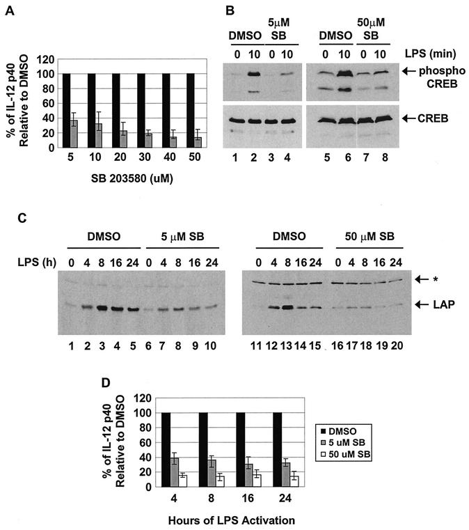
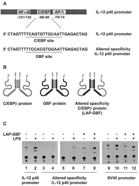
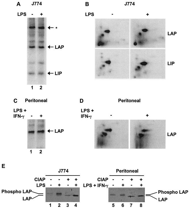
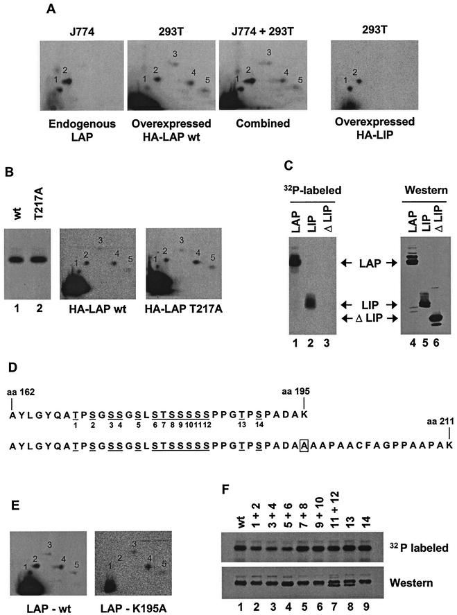
C/EBP family members contribute to the induction of the interleukin-12 p40 gene and the genes encoding several other mediators of inflammation. Here, we show by chromatin immunoprecipitation that C/EBPbeta binds the p40 promoter following lipopolysaccharide stimulation of peritoneal macrophages. However, three modes of C/EBPbeta regulation reported in other cell types were not detected, including alternative translation initiation, nuclear translocation, and increased DNA binding following posttranslational modification. In contrast, C/EBPbeta concentrations greatly increased following stimulation via MAP kinase-dependent induction of C/EBPbeta gene transcription. Increased C/EBPbeta concentrations were unimportant for p40 induction, however, as transcription of the p40 gene initiated before C/EBPbeta concentrations increased. Furthermore, disruption of C/EBPbeta upregulation by a MAP kinase inhibitor only slightly diminished p40 induction. Phosphopeptide mapping revealed that endogenous C/EBPbeta in macrophages is phosphorylated on only a single tryptic peptide containing 14 potential phosphoacceptors. This peptide was constitutively phosphorylated in primary and transformed macrophages, in contrast to its inducible phosphorylation in other cell types in response to Ras and growth hormone signaling. Altered-specificity experiments supported the hypothesis that C/EBPbeta activity in macrophages does not require an inducible posttranslational modification. These findings suggest that, although C/EBPbeta contributes to the induction of numerous proinflammatory genes, it is fully active in unstimulated macrophages and poised to stimulate transcription in conjunction with other factors whose activities are induced.


Figures

FIG. 1.
FIG. 1.
C/EBPβ associates with the IL-12 p40 promoter in LPS-stimulated macrophages. ChIP assays were performed with cross-linked chromatin from wild-type peritoneal macrophages that were unstimulated (−) (lanes 1, 3, and 5) or stimulated (+) with LPS plus IFN-γ for 1 or 4 h (lanes 2, 4, and 6). Immunoprecipitations were performed with GST control antibodies (Ab) (lanes 1 and 2) or C/EBPβ antibodies (lanes 3 and 4). Precipitation of IL-12 p40 and TdT promoter fragments was monitored by PCR. Input samples are in lanes 5 and 6.
FIG.2.
FIG.2.
C/EBPβ protein and DNA-binding activity are upregulated in LPS-stimulated J774 cells. (A) C/EBPβ mRNA was detected by primer extension using 30 μg of total RNA from unstimulated (−) or LPS-stimulated (4 h) (+) J774 cells. (B) C/EBPβ isoforms Full, LAP, and LIP were detected by Western blotting using 60 μg of cytoplasmic (cyto) and nuclear (nuc) extracts from unstimulated or LPS-stimulated (4 h) J774 cells (lanes 1 to 5). Also shown are in vitro-translated C/EBPβ isoforms (lanes 6 to 8). The arrows indicate the locations of LAP, LIP, and a nonspecific protein recognized by the antibody (*). (C) Gel shift and antibody (Ab) supershift assays were performed with a radiolabeled probe containing the C/EBP site from the IL-12 p40 promoter and nuclear extracts from unstimulated or LPS-stimulated (4 h) J774 cells. Binding reactions were performed with no antibody or 2 μg of antibody against C/EBPα, -β, -δ, or -ɛ. (D) A gel shift assay was performed with nuclear extracts from unstimulated or LPS-stimulated (4 h) J774 cells (lanes 1 and 2). As markers, the LAP and LIP isoforms were translated in vitro separately to allow formation of LAP/LAP and LIP/LIP homodimers and were then mixed (lane 3). LAP and LIP were also cotranslated to allow the formation of LAP/LIP heterodimers, as indicated (lane 4). (E) Western blot and gel shift assays were performed with nuclear extracts from unstimulated or LPS-stimulated (4 h) J774 cells. CHX was added (+) to the cells where indicated. (F) RAW264.7 cells were transfected with a CAT reporter plasmid driven by either a constitutive cytomegalovirus (CMV) promoter (lanes 1 and 2), a trimer of the IL-12 p40 C/EBP site upstream of a TATA-Inr core promoter (lanes 3 and 4), or a C/EBPβ promoter (lanes 5 and 6). Transfected cells were left unstimulated or were stimulated with LPS for 24 h. The cells were then harvested, and CAT assays were performed. (G) Whole-cell extracts (100 μg) from unstimulated or LPS-stimulated J774 cells were analyzed by Western blotting using antibodies that recognize phosphorylated CREB (top) or total CREB (bottom).
FIG. 3.
FIG. 3.
Time course of C/EBPβ association and IL-12 p40 expression. (A) Primary p40 transcripts and GAPDH mRNA were monitored by intron PCR in unstimulated or LPS-plus-IFN-γ-stimulated peritoneal macrophages. (B) p40 mRNA was monitored by primer extension in LPS-stimulated J774 cells. (C) p40 mRNA was monitored by primer extension in LPS-plus-IFN-γ-stimulated peritoneal macrophages. (D) p40 protein was monitored by ELISA in supernatants from LPS-stimulated J774 cells. (E) p40 protein was monitored by ELISA in supernatants from LPS-plus-IFN-γ-stimulated peritoneal macrophages.
FIG. 4.
FIG. 4.
Time courses of C/EBPβ upregulation. (A) C/EBPβ mRNA from LPS-stimulated J774 cells was monitored by primer extension. (B) C/EBPβ mRNA from LPS-plus-IFN-γ-stimulated peritoneal macrophages was monitored by primer extension. (C) C/EBPβ isoforms (LAP and LIP) in nuclear extracts (60 μg) from LPS-stimulated J774 cells were monitored by Western blotting. A protein that cross-reacts with the antibody is indicated (*). (D) C/EBPβ isoforms (LAP and LIP) in nuclear extracts (60 μg) from LPS-plus-IFN-γ-stimulated peritoneal macrophages were monitored by Western blotting. (E) C/EBPβ DNA-binding activity was monitored by gel shift, using nuclear extracts (6 μg) from LPS-stimulated J774 cells.
FIG. 5.
FIG. 5.
C/EBPβ is unlikely to be a relevant target of p38 MAP kinase during IL-12 p40 induction. (A) J774 cells were treated for 1 h with different concentrations of SB203580 (shaded bars) or an equivalent volume of the solvent dimethyl sulfoxide (DMSO) (solid bars). The cells were then stimulated with LPS (24 h) in the presence of inhibitor, and p40 protein secretion was monitored by ELISA. The results represent the means (± standard deviations) from three separate experiments. (B) J774 cells were treated for 1 h with different concentrations of SB203580 (SB) or an equivalent volume of DMSO. Phosphorylated CREB and total CREB were then monitored by Western blotting in whole-cell lysates (100 μg) from unstimulated or LPS-stimulated (10 min) cells. (C) J774 cells were treated for 1 h with different concentrations of SB203580 or an equivalent volume of DMSO. LAP was then monitored by Western blotting in nuclear extracts (60 μg) from unstimulated or LPS-stimulated C/EBPβ cells. (D) J774 cells were treated for 1 h with SB203580 or an equivalent volume of DMSO. IL-12 p40 protein was monitored in culture supernatants from LPS-stimulated cells. The data are expressed as percentages of p40 production in DMSO-treated cells.
FIG. 6.
FIG. 6.
Use of an altered-specificity assay to monitor the potential role of C/EBPβ posttranslational modifications. (A) Sequence of the p40 promoter encompassing the C/EBP site. Also shown is the same sequence with the C/EBP site replaced by a GBF binding site. (B) Schematic representations of the C/EBPβ protein, GBF protein, and LAP-GBF fusion protein. (C) RAW264.7 cells were transfected with a CAT reporter plasmid containing the wild-type p40 promoter (−350 to +55; lanes 1 and 2), the altered-specificity p40 promoter (lanes 3 to 8), or a simian virus 40 (SV40) promoter (lanes 9 to 12) either alone (lanes 1 to 6, 9, and 10) or with (+) the LAP-GBF fusion protein (lanes 7, 8, 11, and 12). CAT assays were performed with extracts from unstimulated (−) or LPS-stimulated (24 h) (+) cells.
FIG. 7.
FIG. 7.
Murine C/EBPβ is constitutively phosphorylated in macrophages. (A) J774 cells were incubated with [32P]orthophosphate for 4 h. Phosphorylation of LAP and LIP from unstimulated (−) and LPS-stimulated (20 min) (+) cells was monitored by SDS-PAGE, followed by transfer to nitrocellulose and exposure to film. (B) LAP and LIP were excised from the membrane, digested with trypsin, spotted on a thin-layer chromatography plate, and separated by electrophoresis in the first dimension and chromatography in the second dimension. (C) Phosphorylation of LAP in unstimulated and LPS-plus-IFN-γ-stimulated (20 min) peritoneal macrophages was monitored as described for panel A. The C/EBPβ antibody used for this experiment does not bind LIP. (D) Phosphopeptide mapping of LAP from peritoneal macrophages was performed as decribed for panel B. (E) Untreated and calf intestine alkaline phosphatase (CIAP)-treated LAP were monitored by Western blotting in extracts from unstimulated or LPS-stimulated (4 h) J774 cells or in extracts from unstimulated or LPS-plus-IFN-γ-stimulated (4 h) peritoneal macrophages.
FIG.8.
FIG.8.
The constitutive C/EBPβ phosphorylation sites are within one tryptic peptide. (A) Phosphopeptide maps generated with endogenous LAP (J774 cells) and overexpressed LAP (293T cells) are shown individually (left-hand two maps) and when the two samples were run together (J774 + 293T). A map of overexpressed LIP (293T cells) is also shown (right). HA, hemagglutinin; wt, wild type. (B) Phosphorylation of wild-type LAP and the T217A mutant LAP in 293T cells was monitored by SDS-PAGE (left) and by two-dimensional phosphopeptide mapping (middle and right). (C) Phosphorylation of LAP, LIP, and ΔLIP in 293T cells was monitored by SDS-PAGE. A Western blot shows the overall abundances of the three proteins. (D) Sequence of tryptic peptide phosphorylated in vivo (top), along with sequence of tryptic peptide created by K195A mutation (bottom). aa, amino acid. Potential phosphoacceptors are underlined and numbered. The box corresponds to the residue altered in LAP-K195A. (E) Phosphopeptide maps of wild-type LAP and the K195A mutant LAP in 293T cells. (F) Phosphorylation of LAP mutants that alter potential phosphoacceptors between residues 162 and 195 was monitored by SDS-PAGE of radiolabeled protein (top) and by Western blotting (bottom). Potential phosphoacceptors altered in each mutant protein are indicated above each lane.

References

    1. Agre, P., P. F. Johnson, and S. L. McKnight. 1989. Cognate DNA binding specificity retained after leucine zipper exchange between GCN4 and C/EBP. Science 246:922-926. - PubMed
    1. Akira, S., H. Isshiki, T. Sugita, O. Tanabe, S. Kinoshita, Y. Nishio, T. Nakajima, T. Hirano, and T. Kishimoto. 1990. A nuclear factor for IL-6 expression (NF-IL6) is a member of a C/EBP family. EMBO J. 9:1897-1906. - PMC - PubMed
    1. Alam, T., M. R. An, and J. Papaconstantinou. 1992. Differential expression of three C/EBP isoforms in multiple tissues during the acute phase response. J. Biol. Chem. 267:5021-5024. - PubMed
    1. An, M. R., C.-C. Hsieh, P. D. Reisner, J. P. Rabek, S. G. Scott, D. T. Kuninger, and J. Papaconstantinou. 1996. Evidence for posttranslational regulation of C/EBPα and C/EBPβ isoform expression during the lipopolysaccharide-mediated acute-phase response. Mol. Cell. Biol. 16:2295-2306. - PMC - PubMed
    1. Berrier, A., G. Siu, and K. Calame. 1998. Transcription of a minimal promoter from the NF-IL6 gene is regulated by CREB/ATF and SP1 proteins in U937 promonocytic cells. J. Immunol. 161:2267-2275. - PubMed

Publication types

MeSH terms

LinkOut - more resources