. 2003 Jul;23(14):4882-91.
doi: 10.1128/MCB.23.14.4882-4891.2003.

Normal reproductive function in InhBP/p120-deficient mice

Affiliations

Normal reproductive function in InhBP/p120-deficient mice

Daniel J Bernard et al. Mol Cell Biol. 2003 Jul.

Abstract

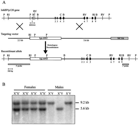
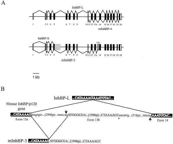
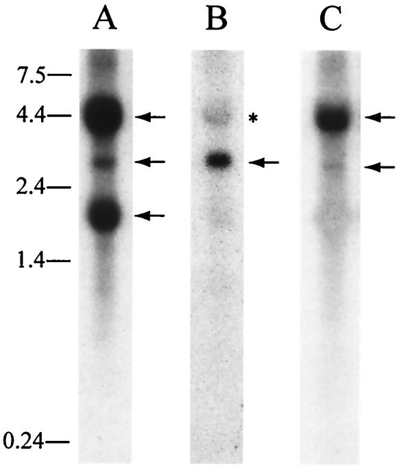
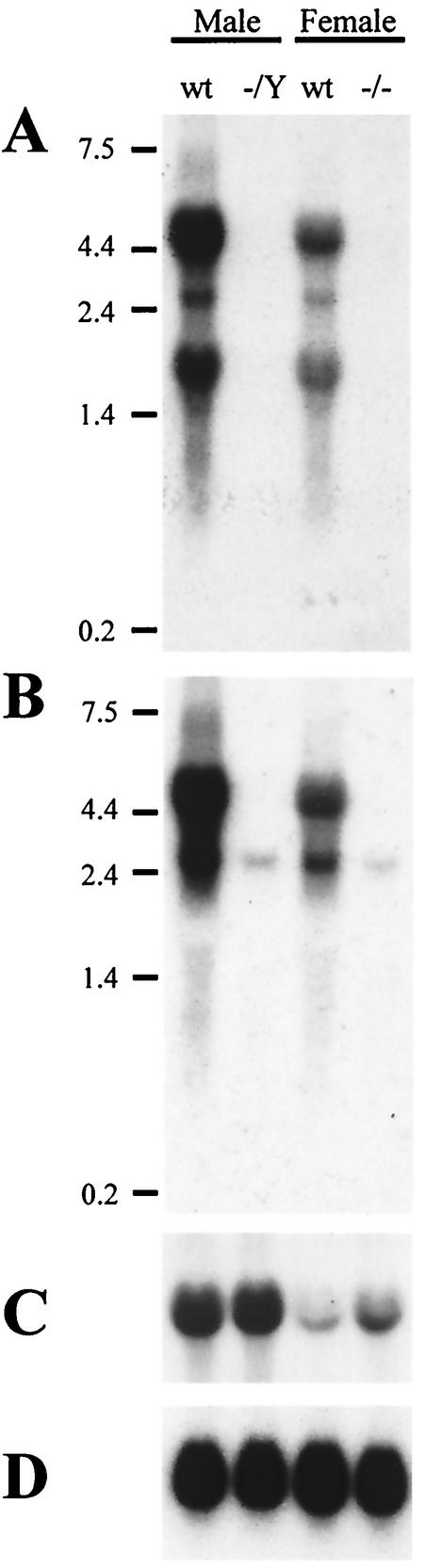
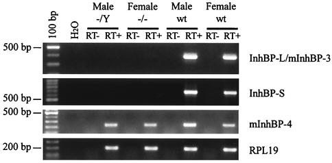
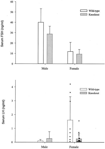
The inhibins are gonadal transforming growth factor beta superfamily protein hormones that suppress pituitary follicle-stimulating hormone (FSH) synthesis. Recently, betaglycan and inhibin binding protein (InhBP/p120, also known as the product of immunoglobulin superfamily gene 1 [IGSF1]) were identified as candidate inhibin coreceptors, shedding light on the molecular basis of how inhibins may affect target cells. Activins, which are structurally related to the inhibins, act within the pituitary to stimulate FSH production. Betaglycan increases the affinity of inhibins for the activin type IIA (ACVR2) receptor, thereby blocking activin binding and signaling through this receptor. InhBP/p120 may not directly bind inhibins but may interact with the activin type IB receptor, ALK4, and participate in inhibin B's antagonism of activin signaling. To better understand the in vivo functions of InhBP/p120, we characterized the InhBP/p120 mRNAs and gene in mice and generated InhBP/p120 mutant mice by gene targeting in embryonic stem cells. InhBP/p120 mutant male and female mice were viable and fertile. Moreover, they showed no alterations in FSH synthesis or secretion or in ovarian or testicular function. These data contribute to a growing body of evidence indicating that InhBP/p120 does not play an essential role in inhibin biology.


Figures

FIG. 1.
FIG. 1.
Generation of InhBP/p120 mutant mice. (A) Schematic representation of the targeting strategy used to generate InhBP/p120-deficient mice. The 428-bp PstI-EcoRV fragment containing exon 1 (and the transcription start site) was replaced with the Pgk-HPRT positive selection cassette. The 5′ and 3′ homology arms are indicated by the thick horizontal lines. Exons appear as boxes and are numbered. Noncoding and coding sequences are indicated in white and black, respectively. The positions of the 5′ internal and 3′ external probes used to screen the ES cells are indicated. B, BamHI; C, ClaI; P, PstI; RI, EcoRI; and RV, EcoRV. (B) Genotyping of offspring was performed by Southern blot analysis of tail genomic DNA digested with EcoRI and EcoRV and hybridized with a cDNA probe corresponding to sequences in exons 2 through 6. The wild-type and recombinant alleles detected in this manner are 5.6 and 9.2 kb, respectively. Note the presence of both alleles in heterozygous females (X+X). Because InhBP/p120 is X-linked, males possess either a wild-type (X+Y) or recombinant (XY) allele.
FIG. 2.
FIG. 2.
Schematic representation of the mouse InhBP/p120 gene and mRNA organization. (A) The mouse InhBP/p120 gene consists of 20 exons and spans 15.2 kb. Exons are numbered and depicted as boxes or vertical lines. Unfilled boxes and filled boxes represent noncoding and coding sequences, respectively. The top of the panel shows the splicing of InhBP-L and mInhBP-4 genes. The former comprises exons 1 through 20, while the latter consists of exons 10 through 20 and is believed to derive from an alternative promoter in intron 9. Both InhBP-L and mInhBP-4 genes use exon 13a. The bottom of the panel shows the splicing of InhBP-S and mInhBP-3 genes. The former comprises exons 1 through 5 as well as part of intron 5 (shaded box). The mInhBP-3 gene comprises exons 1 through 13a plus exon 13b (grey box) at its 3′ end. (B) Schematic representation of the splicing events that give rise to either InhBP-L (top) or mInhBP-3 (bottom). The 3′ end of exon 13a and 5′ end of exon 14 are indicated in white type with black shading. Exon 13b is highlighted with grey shading. In the InhBP-L transcript, the 3′ splice acceptor (ag) at the end of intron 13 (indicated by the upward-pointing arrow) is used. In the mInhBP-3 transcript, an alternative 3′ splice acceptor within intron 13 (indicated by the downward-pointing arrow) is used. The asterisk marks the 3′ end of the mInhBP-3 sequence, where the poly(A) tail begins. Intronic sequences are indicated in lowercase.
FIG. 3.
FIG. 3.
InhBP/p120 transcripts in mouse pituitary gland. Northern blot analyses indicate the presence of four major InhBP/p120 RNA species in adult mouse pituitary. (A) A probe directed against the open reading frame of the rat InhBP-S gene detected transcripts of 4.4 (InhBP-L, top arrow), 2.7 (mInhBP-3, middle arrow), and 1.8 (InhBP-S, bottom arrow) kb. (B) A 204-bp probe corresponding to exon 13b detected a transcript of 2.7 kb (mInhBP-3, arrow). The asterisk indicates the InhBP-L transcript that was incompletely stripped from a previous hybridization. (C) A probe directed against 3′ sequences in exons 18 through 20 detected transcripts of 4.4 (InhBP-L, top arrow) and 2.7 (mInhBP-4, bottom arrow) kb. RNA molecular size standards (in kilobases) are indicated at the left of the figure.
FIG. 4.
FIG. 4.
InhBP/p120 expression in wild-type (wt) and knockout pituitaries. (A) A Northern blot shows expression of InhBP-L (4.4 kb), mInhBP-3 (2.7 kb), and InhBP-S (1.8 kb) in wild-type, but not mutant, mouse pituitaries of males (−/Y) and females (−/−). (B) The same blot hybridized with a 3′ probe shows expression of InhBP-L (4.4 kb) in wild-type, but not knockout, mice. This probe also detected a minor mInhBP-4 band (2.7 kb) in both genotypes. (C) FSHβ mRNA levels were higher in males than in females of both genotypes. The mutation did not significantly affect FSHβ expression in knockout mice relative to that in wild-type animals. (D) Probing of the blot with a rat RPL19 cDNA indicated equal loading of RNA in all lanes.
FIG. 5.
FIG. 5.
RT-PCR analysis of InhBP/p120 isoform expression in mouse pituitaries. RT-PCR analysis confirms the lack of InhBP-L, mInhBP-3, and InhBP-S expression in mutant male and female pituitary glands (top two panels). mInhBP-4 is expressed in males and females of both genotypes (third panel). RPL19 mRNA levels are similar in wild-type and mutant mice (bottom panel). No bands were detected in the H2O only or RT samples. wt, wild-type.
FIG. 6.
FIG. 6.
Serum gonadotropin levels. Radioimmunoassays for FSH (top) and LH (bottom) indicated that serum gonadotropin levels were not significantly affected by the mutation (data reflect the means ± standard deviations). Note that in the LH graph, individual data points are presented for females (black dots). While the two groups do not differ significantly, the large variability observed in the female wild-type group appears to result from elevated LH levels in three animals. An LH surge or pulse was observed in only one of the knockout females. The animals used in these studies were randomly cycling, and these data reflect the normal variability observed when animals are sampled in this fashion.

References

    1. Asashima, M., K. Kinoshita, T. Ariizumi, and G. M. Malacinski. 1999. Role of activin and other peptide growth factors in body patterning in the early amphibian embryo. Int. Rev. Cytol. 191:1-52. - PubMed
    1. Attisano, L., and J. L. Wrana. 2002. Signal transduction by the TGF-beta superfamily. Science 296:1646-1647. - PubMed
    1. Attisano, L., J. L. Wrana, S. Cheifetz, and J. Massague. 1992. Novel activin receptors: distinct genes and alternative mRNA splicing generate a repertoire of serine/threonine kinase receptors. Cell 68:97-108. - PubMed
    1. Attisano, L., J. L. Wrana, E. Montalvo, and J. Massague. 1996. Activation of signalling by the activin receptor complex. Mol. Cell. Biol. 16:1066-1073. - PMC - PubMed
    1. Bernard, D. J., S. C. Chapman, and T. K. Woodruff. 2002. Inhibin binding protein (InhBP/p120), betaglycan, and the continuing search for the inhibin receptor. Mol. Endocrinol. 16:207-212. - PubMed

Publication types

MeSH terms