. 2003 Jul;23(14):5018-30.
doi: 10.1128/MCB.23.14.5018-5030.2003.

Mitotic exit regulation through distinct domains within the protein kinase Cdc15

Affiliations

Mitotic exit regulation through distinct domains within the protein kinase Cdc15

Allison J Bardin et al. Mol Cell Biol. 2003 Jul.

Abstract

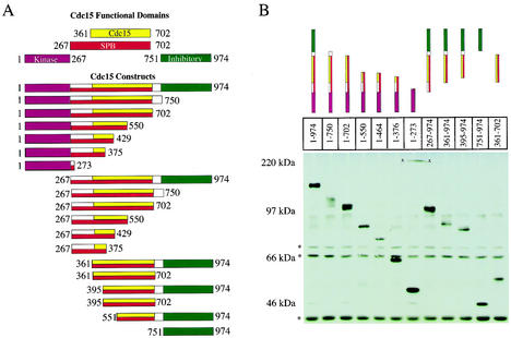
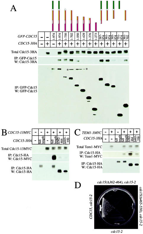
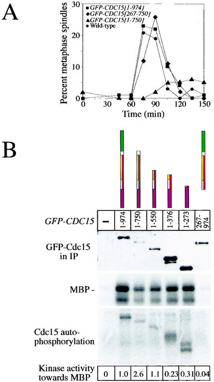
The mitotic exit network (MEN), a Ras-like signaling cascade, promotes the release of the protein phosphatase Cdc14 from the nucleolus and is essential for cells to exit from mitosis in Saccharomyces cerevisiae. We have characterized the functional domains of one of the MEN components, the protein kinase Cdc15, and investigated the role of these domains in mitotic exit. We show that a region adjacent to Cdc15's kinase domain is required for self-association and for binding to spindle pole bodies and that this domain is essential for CDC15 function. Furthermore, we find that overexpression of CDC15 lacking the C-terminal 224 amino acids results in hyperactivation of MEN and premature release of Cdc14 from the nucleolus, suggesting that this domain within Cdc15 functions to inhibit MEN signaling. Our findings indicate that multiple modes of MEN regulation occur through the protein kinase Cdc15.


Figures

FIG. 1.
FIG. 1.
Functional domains of Cdc15 and expression levels of various truncation constructs. (A) The N-terminal kinase domain (aa 1 to 267) is shown in purple; the self-association domain (aa 361 to 702) is shown in yellow; the SPB localization domain (aa 267 to 702) is shown in red, and the inhibitory domain (aa 751 to 974) is shown in green. (B) Anti-GFP Western blot analysis from strains grown in YEP plus 2% raffinose and induced with 2% galactose for 2.5 h. (*, cross-reacting bands; x, nonspecific mark on membrane). The following strains were used: A4683, A5575, A5576, A4692, A5578, A4678, A4690, A4685, A5464, A5466, A6456, and A5909.
FIG. 2.
FIG. 2.
The aa 267 to 702 of Cdc15 are sufficient for SPB localization. (A) Cells were grown to mid-log phase in YEP plus 2% raffinose and induced for 2.5 h with 2% galactose, and Cdc15-GFP was examined in cells producing GFP-Cdc15 [1-974, full-length] (A4441), GFP-Cdc15 [267-974] (A4376), GFP-Cdc15 [267-750] (A4403), or GFP-Cdc15 [361-974] (A5254). (B) Cells were grown to mid-log phase in YEP plus 2% raffinose. Galactose was then added, and the accumulation of GFP-Cdc15 [1-974, full-length] (A4683) and GFP-Cdc15 [361-974] (A5464) was determined at the indicated times. (C) Cells of genotypes described for panel B were induced with galactose for 4 h, and the localization of GFP-Cdc15 full-length and GFP-Cdc15 [361-974] was determined.
FIG. 3.
FIG. 3.
Cdc15 contains a domain that mediates self-association. (A) Cells were grown in YEP plus 2% raffinose and induced with 2% galactose for 2.5 h, and GFP-Cdc15 constructs were immunoprecipitated to determine the amount of associated Cdc15-3HA. (Top) Amount of Cdc15-3HA in 50 μg of whole-cell extracts; (middle and bottom) amount of Cdc15-3HA immunoprecipitated with GFP-Cdc15 and amount of immunoprecipitated GFP-Cdc15 construct, respectively, each from 500 μg of cell extract (*, nonspecific mark on membrane). The following strains were used: A2587, A3932, A4441, A4683, A5575, A5576, A4692, A5578, A4678, A4690, A4685, A5464, A5466, A6456, and A5909. (B) Loss of aa 362 to 464 or 465 to 550 disrupts the ability of Cdc15-3HA to immunoprecipitate Cdc15-13MYC. Cells were grown in yeast extract-peptone-dextrose to mid-log phase, and Cdc15-3HA was immunoprecipitated. (Top) Amount of Cdc15-13MYC in extracts; (middle) amount of Cdc15-13MYC immunoprecipitated with Cdc15-3HA; (bottom) amount of immunoprecipitated Cdc15-3HA. The following strains were used: A2587, A3932, A5401, A6501, A6502, and A6503. (C) Loss of self-association domains does not affect the ability of Cdc15 to interact with Tem1. Cells were grown in yeast extract-peptone-dextrose to mid-log phase, and Cdc15-3HA was immunoprecipitated. (Top) Amount of Tem1-3MYC in extracts; (middle) amount of Tem1-3MYC immunoprecipitated with Cdc15-3HA; (bottom) amount of immunoprecipitated Cdc15-3HA. The following strains were used: A2587, A1828, A6542, A6543, and A6544. (D) The self-association domain is essential for viability. Strains expressing either wild-type CDC15 (A6546 [cdc15-2 CDC15-3HA]) or CDC15 lacking amino acids required for self-association (A6547 [cdc15-2 CDC15(Δ362-464-3HA)] and A6548 [cdc15-2 CDC15(Δ465-550-3HA)]) were streaked to single colonies on yeast extract-peptone-dextrose plates and grown at 37°C for 36 h.
FIG. 4.
FIG. 4.
Overexpression of full-length GFP-CDC15 does not affect associated kinase activity of Cdc15-3HA. Cells were grown in YEP plus 2% raffinose overnight to mid-log phase. Cultures were diluted to an optical density at 600 nm of 0.2 and arrested in metaphase with 15 μg of nocodazole/ml for 1.5 h. An additional 15 μg of nocodazole/ml was added to prevent cells from escaping the metaphase arrest, and cells were induced for 2.5 h with 2% galactose (except lane 3, which is uninduced). The amount of kinase activity associated with Cdc15-3HA was assayed by immunoprecipitation followed by a kinase assay with MBP as an artificial substrate. The following strains were used: A2587, A3932, A4683, A4686, and A4678.
FIG. 5.
FIG. 5.
Several GFP-CDC15 truncation constructs cause a telophase arrest upon overexpression. Strains were arrested for 3 h in G1 with the α-factor pheromone (5 μg/ml) and induced with 2% galactose for the last hour. Cells were synchronously released into YEP plus 2% raffinose and 2% galactose, and samples were taken at indicated time points. (A) The key at right indicates strains A3932, A4441, and A4376 (top to bottom, respectively). Dashed lines represent metaphase spindles; solid lines represent anaphase/telophase spindles. (B) Growth conditions were as described for panel A, and the percentages of anaphase and telophase spindles are analyzed. The leftmost panel depicts class 1 Cdc15 constructs. The key shows strains A3932, A5574, A4695, A5576, A5051, A4691, and A4687 (top to bottom, respectively). Class 2 Cdc15 constructs are shown in the middle panel. The key shows strains A3932, A5580, A4680, A4677, and A6373 (top to bottom, respectively). The rightmost panel depicts class 3 Cdc15 constructs. The key shows strains A3932, A5464, A5909, A5466, A5914, A6456, and A4690 (top to bottom, respectively).
FIG. 6.
FIG. 6.
Expression of Cdc15 lacking the C terminus causes a delay in entry into mitosis and an increase in specific kinase activity. (A) Strains were arrested for 3 h in G1 with the α-factor pheromone (5 μg/ml) and induced with 2% galactose for the last hour. Cells were then synchronously released into YEP plus 2% raffinose and 2% galactose, and the percentage of cells with metaphase spindles was determined at the indicated time. Circles, strain A3932; squares, strain A4441; triangles, strain A5574; and diamonds, strain A4695. (B) Cultures were prepared as described in the legend to Fig. 4, GFP-Cdc15 proteins were immunoprecipitated, and associated kinase activity was measured with MBP as an artificial substrate. (Top) Western blot analysis of the amount of GFP-Cdc15 proteins present in immunoprecipitates used for kinase assays; (middle) amount of 32P-labeled MBP from in vitro kinase assays; (bottom) autophosphorylation occurring on immunoprecipitated GFP-Cdc15. The amount of 32P-labeled MBP was determined relative to the amount of GFP-Cdc15 protein present in the immunoprecipitates. The following strains were used: A3932, A4683, A5575, A4692, A4678, A4690, and A4686.
FIG. 7.
FIG. 7.
Removal of a C-terminal domain of Cdc15 causes hyperactivation of MEN. (A) Strains were arrested for 3 h in G1 with the α-factor pheromone (5 μg/ml) and induced with 2% galactose for the last hour. Cells were synchronously released into YEP plus 2% raffinose and 2% galactose, and Dbf2 kinase activity and the amount of Dbf2 and Cdc28 (loading control) protein were determined at the indicated times. The following strains were used: A6131, A6130, A5568, and A5571. (B and C) Strains were grown to mid-log phase in YEP plus 2% raffinose and arrested in metaphase with 15 μg of nocodazole/ml for 1.5 h. An additional 7.5 μg of nocodazole/ml was added to maintain the metaphase arrest, and expression of GFP-CDC15 constructs was induced with 2% galactose. Samples were taken at indicated time points to determine the percentage of cells with Cdc14 released from the nucleolus. The image shown in panel C shows Cdc14 localization 90 min after galactose addition. Cdc14 staining is shown in red, and DNA is shown in blue. The following strains were used: A1411, A5965, A5966, and A5968.

Similar articles

Cited by

References

    1. Asakawa, K., S. Yoshida, F. Otake, and A. Toh-e. 2001. A novel functional domain of Cdc15 kinase is required for its interaction with Tem1 GTPase in Saccharomyces cerevisiae. Genetics 157:1437-1450. - PMC - PubMed
    1. Bardin, A. J., and A. Amon. 2001. Men and sin: what's the difference? Nat. Rev. Mol. Cell Biol. 2:815-826. - PubMed
    1. Bardin, A. J., R. Visintin, and A. Amon. 2000. A mechanism for coupling exit from mitosis to partitioning of the nucleus. Cell 102:21-31. - PubMed
    1. Cenamor, R., J. Jimenez, V. J. Cid, C. Nombela, and M. Sanchez. 1999. The budding yeast Cdc15 localizes to the spindle pole body in a cell-cycle-dependent manner. Mol. Cell Biol. Res. Commun. 2:178-184. - PubMed
    1. Dan, I., N. M. Watanabe, and A. Kusumi. 2001. The Ste20 group kinases as regulators of MAP kinase cascades. Trends Cell Biol. 11:220-230. - PubMed

Publication types

MeSH terms