. 2003 Jun 15;23(12):4793-7.
doi: 10.1523/JNEUROSCI.23-12-04793.2003.

Calcium signaling in single peripheral sensory nerve terminals

Affiliations

Calcium signaling in single peripheral sensory nerve terminals

Tony D Gover et al. J Neurosci. .

Abstract

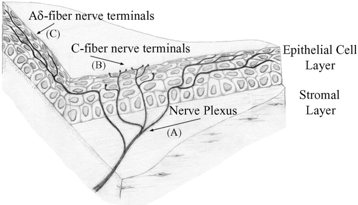
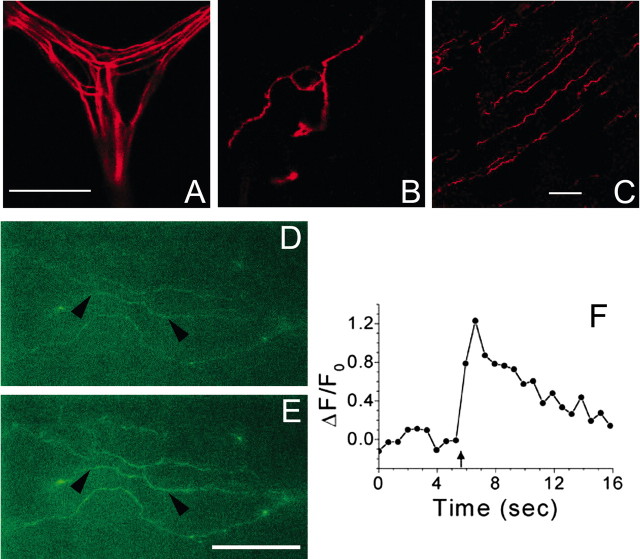
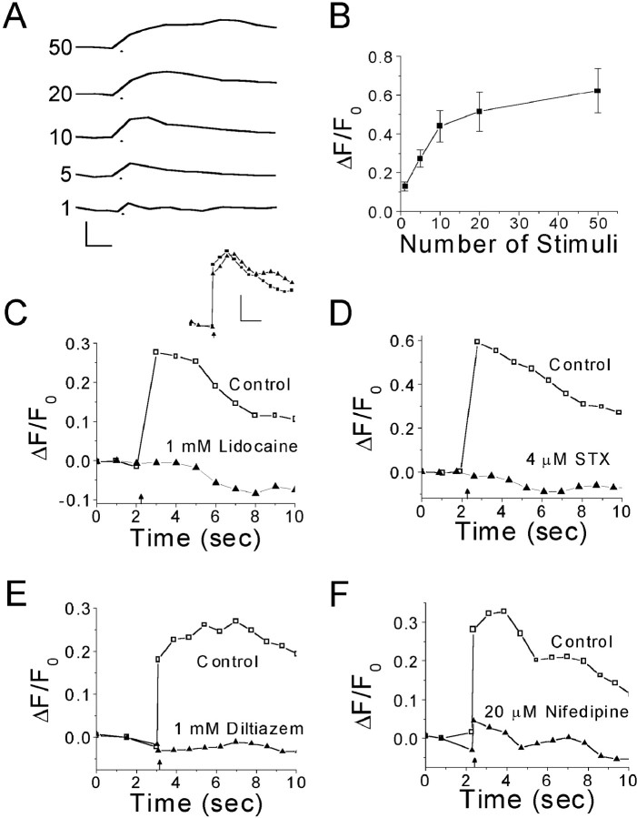
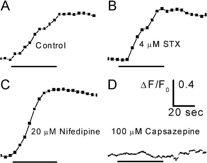
Peripheral sensory nerve terminals (PSNTs) have a dual function: reporting normal and abnormal sensations and releasing trophic factors to maintain the structure and function of epithelial cells. Although it is widely considered that intracellular Ca2+ plays a critical signaling role for both functions, the role of Ca2+ signaling has never been studied in PSNTs, primarily because of their small size and anatomical inaccessibility. Here, using epifluoresence microscopy and a fluorescent Ca2+ indicator, we report that action potentials or chemical irritation can elicit transient rises in [Ca2+]i (Ca2+ transients) in PSNTs within the corneal epithelium of the rat. In vitro electrical stimulation of the ciliary nerves in the eye, or electrical field stimulation of the cornea, evoked Ca2+ transients with a magnitude that was proportional to the number of stimuli applied over the range of 1-10 stimuli. Ca2+ transients were significantly blocked by 1 mm lidocaine, 4.1 microm saxitoxin (STX), or L-type Ca2+ channel antagonists (1 mm diltiazem or 20 microm nifedipine). The nociceptive agonist capsaicin (1 microm) elicited Ca2+ transients in all nerve terminals studied. Capsaicin-evoked Ca2+ transients were completely blocked by the vanilloid receptor 1 antagonist capsazepine (100 microm). In contrast, capsaicin-evoked Ca2+ transients were not attenuated by preincubation with 4.1 microm STX or 20 microm nifedipine. These findings demonstrate, for the first time, that nerve impulses or chemical stimulation promote Ca2+ entry into PSNTs, including nociceptors.


Figures

Figure 1.
Figure 1.
Illustration of the branching pattern of Aδ and C fiber nerve terminals within the cornea. Aδ and C fibers course together in the collagenous stromal layer. The nerve plexus in the stromal layer consists of extensively branching Aδ and C fibers. The fibers separate as they turn toward the superficial epithelial layers, where they terminate. Letters in parentheses correspond to the images in Figure 2. Adapted from MacIver and Tanelian (1993).
Figure 2.
Figure 2.
Fluorescent labeling of corneal nerve fibers. A–C, Nerve fibers were loaded with tetramethylrhodamine dextran (10 kDa) 24 hr before confocal microscopy. A, Subepithelial nerve plexus lying within the stromal layer. B, Superficial nerve terminals ∼5 μm below corneal surface. C, Nerve terminals lying deeper within the corneal epithelium. Scale bars: (in A) A, B, 25 μm; C, 50 μm. D, E, Nerve terminals loaded with Oregon Green 488 BAPTA-1 dextran (10 kDa) 24 hr before imaging with a cooled CCD camera. D, Nerve terminals imaged immediately before electrical stimulation. E, Same nerve terminals as in D imaged 1 sec after the start of a train of 10 field stimuli (10 Hz). F, ΔF/F0 trace for a section (marked by arrowheads) of the middle nerve terminal seen in D and E; arrow indicates the start of electrical stimulation. Scale bar: (in E) D, E, 35 μm. For Ca2+ imaging in D—F, 500 msec exposures were collected at 1.67 Hz.
Figure 3.
Figure 3.
Effect of Na+ and Ca2+ channel antagonists on Ca2+ transients evoked by electrical stimulation in corneal nerve terminals. A, Average Ca2+ transients evoked by electrical stimulation of the ciliary nerves. The number of stimuli (10 Hz) used to evoke a Ca2+ transient is indicated next to each trace. The dot below each trace marks the start of stimulation. Trace shown for one stimulus is the average of recordings from five different nerve terminals; images were 100 msec exposures captured at 2.5 Hz. For 5–50 stimuli, records from seven separate terminals were averaged; images were 400 msec exposures acquired at 1.4 Hz. Calibration: 0.5 ΔF/F0, 1.0 sec. B, Plot of average peak ΔF/F0 from A against number of stimuli. Note the nonlinearity in the relationship between peak ΔF/F0 and the number of stimuli. The plateau was not caused by indicator or detector saturation (see Discussion and Appendix). Error bars indicate ±SEM. C, Ca2+ transients evoked in a single nerve terminal by antidromic stimulation before and after 20 min perfusion with 1 mm lidocaine. Inset, Control records of Ca2+ transients evoked by antidromic stimulation before (squares) and after (triangles) 20 min perfusion of Locke solution containing no drug. Calibration: 0.2 ΔF/F0, 2.0 sec. D,Ca2+ transients evoked in a single nerve terminal by antidromic stimulation before and after 30 min perfusion with 4.1 μm STX. E, Ca2+ transients evoked in a single nerve terminal by field stimulation before and after 20 min perfusion with 1 mm diltiazem. F, Ca2+ transients evoked in a single nerve terminal by antidromic stimulation before and after 20 min perfusion with 20 μm nifedipine. In C—F, the arrow marks the start of electrical stimulation (20 stimuli at 10 Hz); data acquired in the absence of antagonist are represented by open symbols on traces labeled Control; data acquired in the presence of antagonist are represented by closed symbols on traces labeled with the antagonist name and concentration; 400 msec exposures were acquired at 1.4 Hz; traces were smoothed by three-point averaging.
Figure 4.
Figure 4.
Ca2+ transients evoked by capsaicin in corneal nerve terminals. A, Response of an individual nerve terminal to 1 μm capsaicin. B, Response of an individual nerve terminal to 1 μm capsaicin after a 30 min preincubation with 4.1 μm STX. C, Response of an individual nerve terminal to 1 μm capsaicin after a 20 min preincubation with 20 μm nifedipine. D, The response to capsaicin was completely blocked by a 35 min preincubation with 100 μm capsazepine, aVR1 antagonist. The time bar under each trace indicates the duration of capsaicin application. Images were 400 msec exposures acquired at 0.2 Hz (A—C) or 1.0 Hz (D). All traces were smoothed by three-point averaging.

References

    1. Belmonte C, Gallar J ( 1996) Corneal nociceptors. In: Neurobiology of nociceptors (Belmonte C, Cervero F, eds), pp 146–183. New York: Oxford UP.
    1. Black JA, Waxman SG ( 2002) Molecular identities of two tetrodotoxin-resistant sodium channels in corneal axons. Exp Eye Res 75: 193–199. - PubMed
    1. Brock JA, Mclachlan EM, Belmonte C ( 1998) Tetrodotoxin-resistant impulses in single nociceptor nerve terminals in guinea-pig cornea. J Physiol (Lond) 512: 211–217. - PMC - PubMed
    1. Caterina MJ, Schumacher MA, Tominaga M, Rosen TA, Levine JD, Julius D ( 1997) The capsaicin receptor: a heat-activated ion channel in the pain pathway. Nature 398: 816–824. - PubMed
    1. Chen X, Belmonte C, Rang HP ( 1997a) Capsaicin and carbon dioxide act by distinct mechanisms on sensory nerve terminals in the cat cornea. Pain 70: 23–29. - PubMed

Publication types

MeSH terms

LinkOut - more resources