. 2003 Jun 15;23(12):4842-9.
doi: 10.1523/JNEUROSCI.23-12-04842.2003.

Early maintenance of hippocampal mossy fiber--long-term potentiation depends on protein and RNA synthesis and presynaptic granule cell integrity

Affiliations

Early maintenance of hippocampal mossy fiber--long-term potentiation depends on protein and RNA synthesis and presynaptic granule cell integrity

Eduardo Calixto et al. J Neurosci. .

Abstract

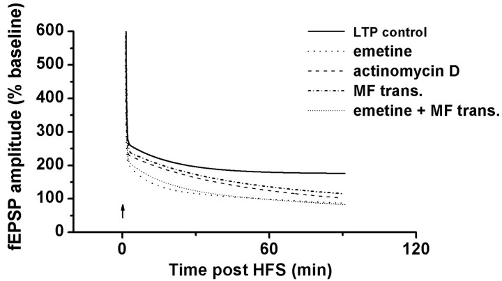
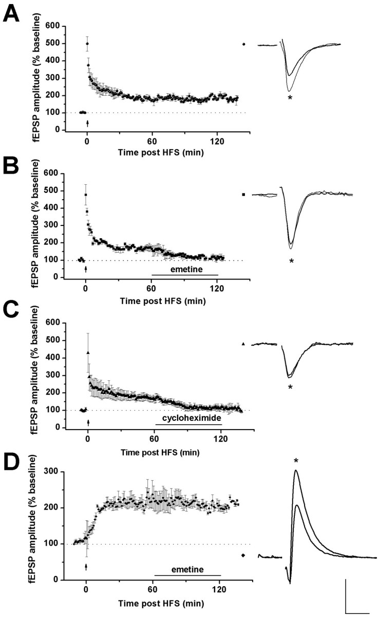
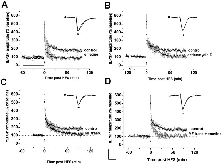
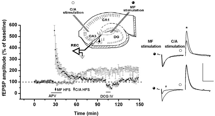
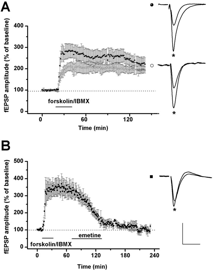
The neural substrates of memory likely include long-term potentiation (LTP) of synaptic strength that results from high-frequency stimulation (HFS) of the afferent pathway. The mechanisms that underlie the maintenance of LTP include RNA and protein synthesis, although the contribution of these molecular events typically does not become essential until several hours after LTP induction. We here show that, different from this pattern, (1) LTP maintenance at the mossy fiber (MF) input to CA3 pyramidal cells in the hippocampus depends on protein and RNA synthesis soon after LTP induction, and (2) some of these molecular events are controlled by signaling from the presynaptic granule cell soma. Bath application of the protein synthesis inhibitor emetine or cycloheximide 1 hr after MF LTP induction in hippocampal slices caused loss of MF potentiation. In contrast, application of emetine 1 hr after LTP induction at the commissural-associational input to CA3 pyramidal cells had no effect on this form of LTP. Administration of emetine or the RNA synthesis inhibitor actinomycin-D before delivery of HFS to MF input also caused a rapid decay of MF potentiation, although neither drug had an effect on the amplitude or the time-constant of decay of post-tetanic potentiation (PTP). Similarly, transection of MF axons near granule cell somas had no effect on baseline or PTP parameters but caused loss of potentiation at a rate comparable with that after actinomycin-D application. These results indicate that the mechanisms that underlie MF LTP maintenance differ from those involved in LTP maintenance at other glutamatergic synapses.


Figures

Figure 3.
Figure 3.
Time course of the decay of MF fEPSP amplitude after HFS. Curves are the mathematical best fits of data from control experiments (Fig. 1 A) and the experiments shown in Figure 2 A—D. The time course of LTP in control experiments was fit best with model A. In this case, PTP decayed to a nondecremental level of LTP (see Materials and Methods). Because of the application of either emetine or actinomycin-D before delivery of HFS, the decay of LTP was fit best with model B (see Materials and Methods). Model B assumes that the amplitude of both PTP and LTP decayed with different time constants back to pre-HFS amplitude values (see Materials and Methods). Upward arrow indicates the time at which HFS was delivered.
Figure 1.
Figure 1.
Inhibition of protein synthesis blocks early maintenance of MF LTP but not of C/A LTP. A, Control experiments. Left panel, Group data (n = 5; mean ± SEM; circles) of the amplitude of MF fEPSPs evoked by stimulation of MF before and after delivery of HFS (upward arrows). After PTP the amplitude of the response decayed gradually to a stable LTP. Right panel, Single traces of MF fEPSPs recorded during baseline and 120 min after the induction of LTP (asterisk). B, Left panel, Group data (n = 3; mean ± SEM; squares) of the effect of emetine (20 μm; applied 60 min after delivery of HFS; upward arrow) on MF LTP. Right panel, Traces are single MF fEPSPs recorded during baseline and at the end of emetine perfusion (asterisk). C, Left panel, Group data (n = 5; mean ± SEM; triangles) of effect of cycloheximide (60 μm; applied 60 min after HFS; upward arrow) on MF LTP. Right traces are single MF fEPSPs recorded during baseline and at the end of cycloheximide perfusion (asterisk). Cycloheximide and emetine significantly reduced MF LTP (fEPSP amplitude before drug application vs during drug application: p < 0.01). All experiments were performed in the presence of MK-801 (15 μm). D, Left panel, Group data of the amplitude of C/A fEPSPs evoked by stimulation of s. radiatum in area CA2. Emetine application (20 μm; rhombus; n = 3) 60 min after the induction of LTP by HFS delivered to C/A fibers (upward arrow) did not affect the potentiated responses. Right panel, Representative traces of C/A fEPSPs before and at the end of emetine perfusion (asterisk). The horizontal bar indicates duration of drug perfusion (60 min). Calibration: 0.2 mV, 10 msec.
Figure 2.
Figure 2.
Early maintenance of MF LTP is blocked by inhibition of either protein or RNA synthesis as well as by MF transection. Group data (mean ± SEM) of the amplitude of MF fEPSPs before and after HFS (upward arrow). The horizontal bar in A, B, and D indicates the duration of drug application before HFS. A, Effect of emetine incubation for 60 min before delivery of HFS to the MF (20 μm; n = 3; triangles). B, Effect of actinomycin-D incubation for 120 min before delivery of HFS to the MF (25 μm; n = 4; filled squares). C, Effect of MF transection performed 120 min before the onset of electrophysiological recordings (n = 5; filled pentagon). The same HFS as delivered in these experiments induced persistent MF LTP in control slices (filled circles; n = 5). D, Joint effect of emetine incubation (20 μm; 60 min before delivery of HFS) and MF transection on MF LTP (n = 4; open squares). Representative traces of MF fEPSPs were obtained 10 min before and 60 min after HFS or 90 min after delivery of HFS in slices with MF transection (asterisk). Calibration: 0.2 mV, 10 msec.
Figure 4.
Figure 4.
MF transection does not affect the ability of CA3 pyramidal cells to maintain LTP induced at a non-MF synaptic input. Left panel, MF fEPSPs were recorded in the presence of APV (50 μm) for 20 min before the delivery of HFS (first upward arrow). Delivery of HFS to MF induced PTP but not MF LTP (filled circles; n = 3). Thirty minutes after delivery of HFS to MF, a second HFS delivered to the C/A fibers (second upward arrow) induced stable C/A LTP (open circles). In the presence of DCG IV (0.5 μm for 15 min), MF fEPSPs were reduced selectively. Insert, Schematic of hippocampal slice showing the position of the stimulation and recording electrodes and the transection of MF. Right panel, Single traces of MF fEPSPs (left) and C/A fEPSPs (right). Top traces show fEPSP before and 30 min after delivery of HFS to the C/A input. Only C/A fEPSPs are potentiated (asterisk). Bottom traces show the selective depression of DCG IV on MF fEPSPs(#). The horizontal bar indicates the duration of drug application. Calibration: 0.2 mV, 10 msec.
Figure 5.
Figure 5.
Forskolin induces potentiation of MF fEPSPs in control and MF transected slices that is dependent on protein synthesis. A, Left panel shows the time course of potentiation after transient application of forskolin (100 μm)/IBMX (50 μm; 20 min, horizontal bar; in the presence of MK 801, 15 μm) in slices with MF transection (n = 4; open circles) or control slices (n = 5; filled circles). MF LTP in slices with MF transection was not statistically different from MF LTP in control slices 30 min after exposure to forskolin/IBMX (p > 0.4). Right panel shows examples of MF fEPSPs recorded during the baseline period and 60 min after forskolin/IBMX perfusion (asterisk); top traces, MF LTP in control slices; bottom traces, fEPSP from slices with MF transection. B, Effect of emetine incubation (20 μm; 60 min) on potentiation induced by forskolin/IBMX (n = 3; filled squares). After emetine application, MF fEPSPs returned to baseline values. Right panel shows MF fEPSPs recorded during baseline and 60 min after termination of emetine incubation (asterisk). Calibration: 0.2 mV, 10 msec.

References

    1. Aakalu G, Smith WB, Nguyen N, Jiang C, Schuman EM ( 2001) Dynamic visualization of local protein synthesis in hippocampal neurons. Neuron 30: 489–502. - PubMed
    1. Barea-Rodríguez EJ, Rivera DT, Jaffe DB, Martínez Jr JL ( 2000) Protein synthesis inhibition blocks the induction of mossy fiber long-term potentiation in vivo. J Neurosci 20: 8528–8532. - PMC - PubMed
    1. Bennet MR ( 2000) The concept of long term potentiation of transmission at synapse. Prog Neurobiol 60: 109–137. - PubMed
    1. Brown A ( 2000) Slow axonal transport: stop and go traffic in the axon. Nat Rev Mol Cell Biol 1: 153–156. - PubMed
    1. Castillo EP, Janz R, Südhof TC, Tzounopoulos T, Malenka RC, Nicoll RA ( 1997) Rab3A is essential for mossy fibre long-term potentiation in the hippocampus. Nature 388: 590–593. - PubMed

Publication types

MeSH terms

LinkOut - more resources