Comparative Study
. 2003 Jun 15;23(12):4858-67.
doi: 10.1523/JNEUROSCI.23-12-04858.2003.

Increased susceptibility of striatal mitochondria to calcium-induced permeability transition

Affiliations
Comparative Study

Increased susceptibility of striatal mitochondria to calcium-induced permeability transition

Nickolay Brustovetsky et al. J Neurosci. .

Abstract

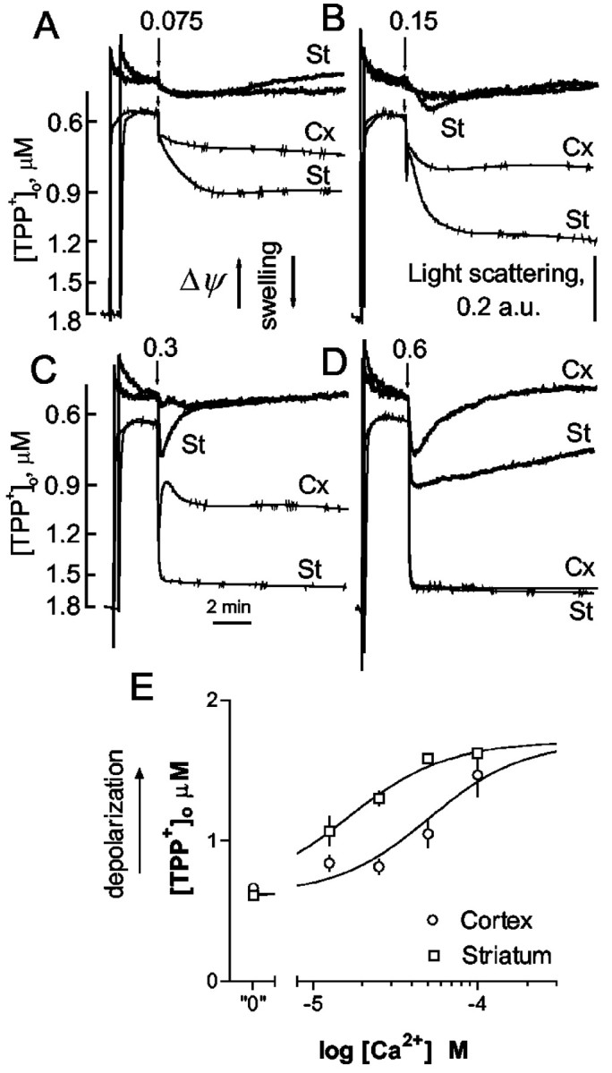
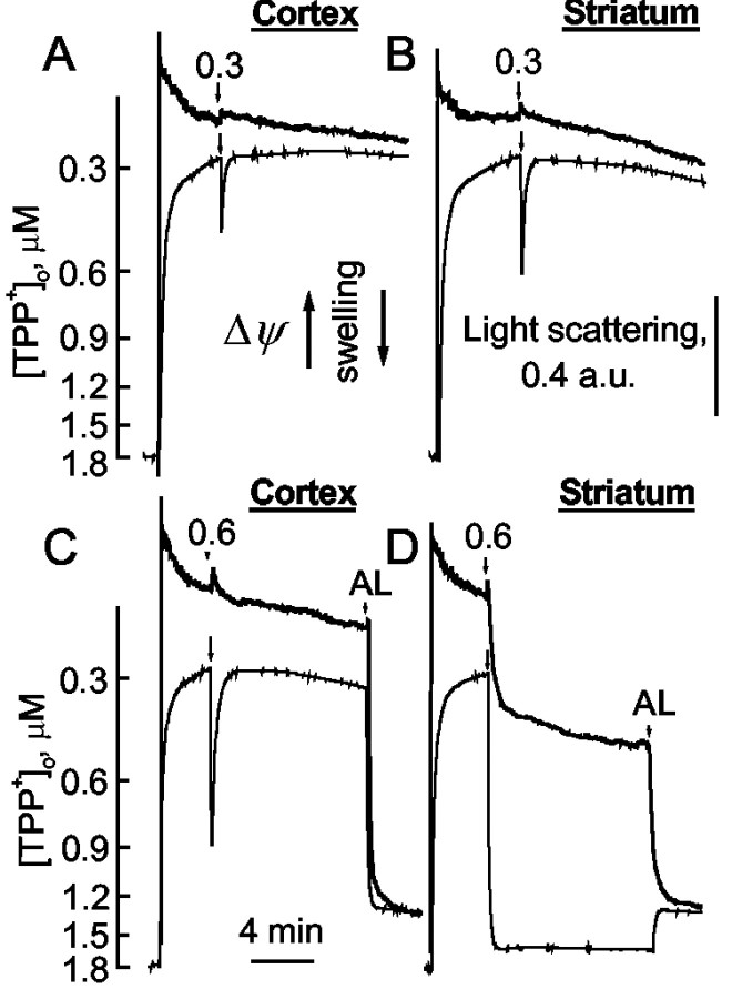
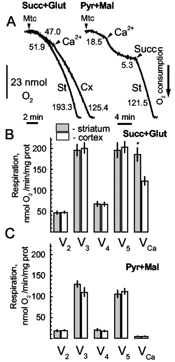
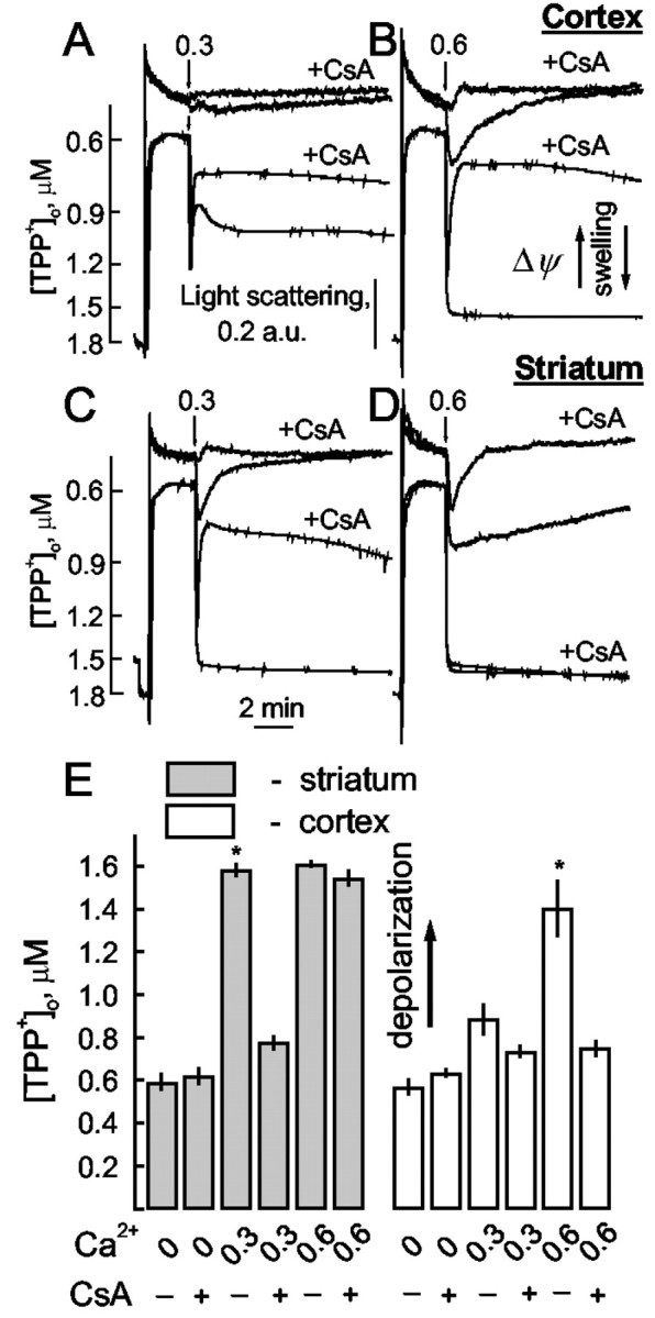
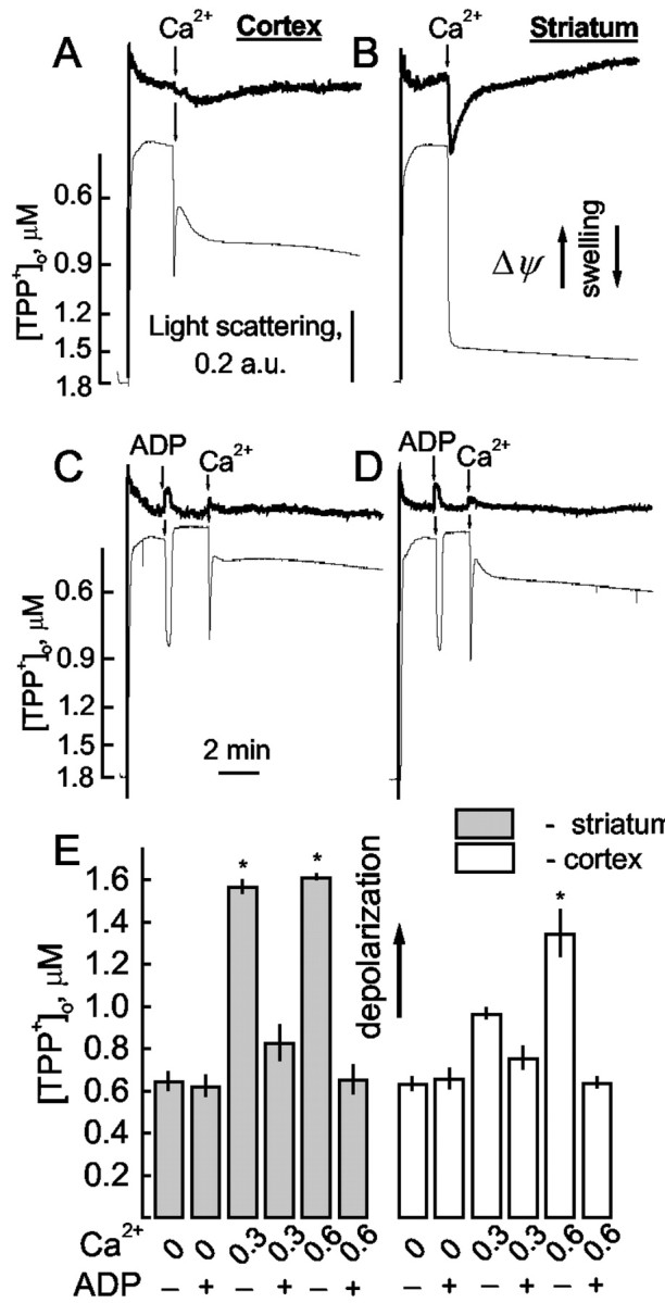
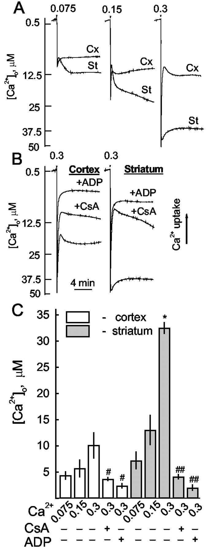
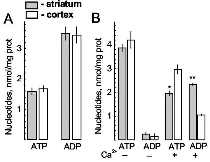
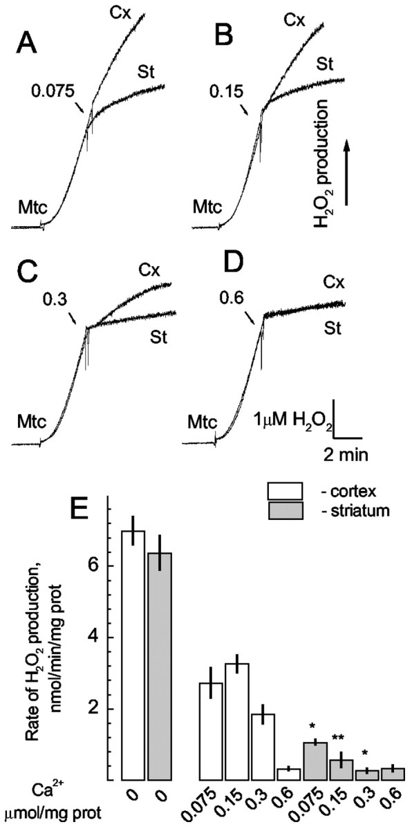
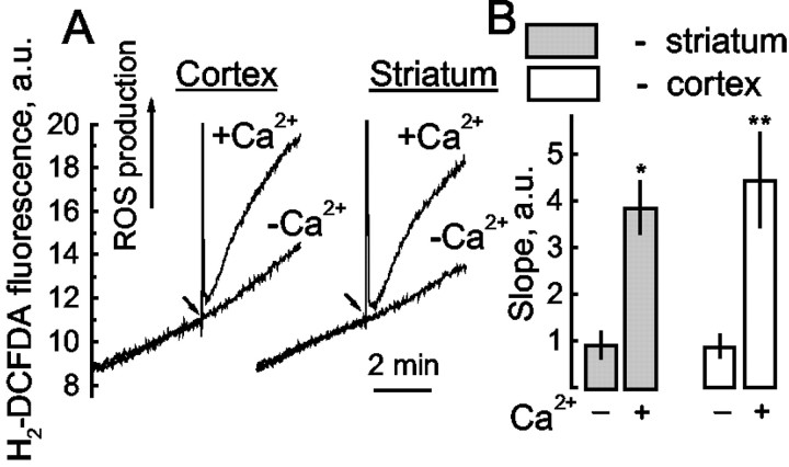
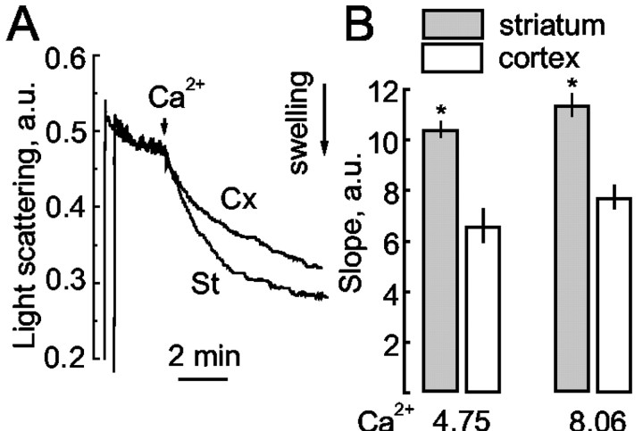
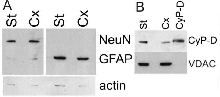
Mitochondria were simultaneously isolated from striatum and cortex of adult rats and compared in functional assays for their sensitivity to calcium activation of the permeability transition. Striatal mitochondria showed an increased dose-dependent sensitivity to Ca2+ compared with cortical mitochondria, as measured by mitochondrial depolarization, swelling, Ca2+ uptake, reactive oxygen species production, and respiration. Ratios of ATP to ADP were lower in striatal mitochondria exposed to calcium despite equal amounts of ADP and ATP under respiring and nonrespiring conditions. The Ca2+-induced changes were inhibited by cyclosporin A or ADP. These responses are consistent with Ca2+ activation of both low and high permeability pathways constituting the mitochondrial permeability transition. In addition to the striatal supersensitivity to induction of the permeability transition, cyclosporin A inhibition was less potent in striatal mitochondria. Immunoblots indicated that striatal mitochondria contained more cyclophilin D than cortical mitochondria. Thus striatal mitochondria may be selectively vulnerable to the permeability transition. Subsequent mitochondrial dysfunction could contribute to the initial toxicity of striatal neurons in Huntington's disease.


Figures

Figure 1.
Figure 1.
Calcium sensitivity of striatal (St) and cortical (Cx) mitochondria in KCl-based medium. AD, Mitochondrial TPP + accumulation (Δψ, thin lines) and light scattering (swelling, thick lines) were monitored in response to the indicated Ca2+ pulses presented in micromoles of Ca2+ per milligram of mitochondrial protein (AD). E, External TPP + concentrations ([TPP +]o) 5 min after Ca2+ addition as a function of added Ca2+. In all figures, numbers and arrows indicate Ca2+ addition in micromoles per milligram of protein. Curves shown are drawn through the averaged data points. Statistical comparisons were made between curves fitted to the data of each individual day.
Figure 2.
Figure 2.
Calcium sensitivity of striatal and cortical mitochondria in mannitol—sucrose medium. Measurements and symbols are as in Figure 1. Alamethicin (AL) (30 μg/ml) produced maximal swelling.
Figure 3.
Figure 3.
Oxygen consumption of cortical and striatal mitochondria respiring on 3 mm succinate and 3 mm glutamate (A, B) or 3 mm pyruvate and 1 mm malate (A, C) in respiratory states 2 (V2, before ADP addition), 3 (V3, after ADP addition), 4 (V4, after ADP depletion), and 5 (V5, after addition of 40 μm 2,4-dinitrophenol) and in response to 0.3 μmol Ca2+/mg protein (VCa2+). Respiratory rates from three to six experiments (A) were averaged and compared in B and C. *p < 0.01 comparing striatal with cortical mitochondria.
Figure 4.
Figure 4.
CsA (1 μm) prevented Ca2+-induced depolarization and swelling in both cortical (A, B) and striatal (C, D) mitochondria, but this inhibition was overcome by lower [Ca2+] in striatal mitochondria (B,D). Traces are the same as in Figure 1. E, Summary of averaged [TPP +]o measurements before or 5 min after Ca2+ addition from three to eight experiments applying 1 μm CsA as antagonist. *p < 0.001 versus measurements with the same Ca2+ in the presence of CsA.
Figure 5.
Figure 5.
ADP antagonized Ca2+-induced depolarization and swelling. Depolarizations and swelling in cortical (A, C) and striatal mitochondria (B, D) were prevented by ADP (C, D). E, Summary of TPP + measurements from three to eight experiments applying ADP as antagonist. Averaged [TPP +]o before or 5 min after Ca2+ addition with or without 100 μm ADP. *p < 0.001 versus measurements with the same Ca2+ in the presence of ADP.
Figure 6.
Figure 6.
Calcium uptake was depressed in striatal (St) compared with cortical (Cx) mitochondria for similar Ca2+ additions (A). CsA (1 μm) or ADP (100 μm) increased Ca2+ uptake to comparable levels in both tissues (B). C, External [Ca2+] 5 min after indicated Ca2+ additions reflects differences in mitochondrial type and the presence of CsA or ADP. *p < 0.001 striatal mitochondria compared with cortical mitochondria. #p < 0.01, ##p < 0.001 compared with Ca2+ addition in the same type of mitochondria in the absence of antagonist.
Figure 7.
Figure 7.
Adenine nucleotide content was comparable between striatal and cortical mitochondria under all conditions except after addition of 0.3 μmol Ca2+/mg protein. AdNs were assayed in mitochondrial suspension (100 μl, 20 μg protein) taken after either storage on ice (A) or incubation in the incubation medium at 37°C (B), before or 5 min after addition of 0.3 μmol Ca2+/mg protein at 37°C. *p < 0.01, **p < 0.001 comparing striatum with cortex.
Figure 8.
Figure 8.
H2O2 production monitored by Amplex Red fluorescence decreased in striatal (St) mitochondria more than in cortical (Cx) mitochondria in response to the indicated doses of Ca2+ (AD). E, Averaged initial slopes of the Amplex Red traces indicate differing rates of H2O2 production. *p < 0.01, **p < 0.001 striatum compared with cortex.
Figure 9.
Figure 9.
ROS generation monitored with H2-DCFDA before and after addition of 0.3 μmol Ca2+/mg protein in cortical and striatal mitochondria (A). B, Comparisons of initial rates of H2-DCFDA fluorescence increase (mean ± SEM) in both types of mitochondria. *p < 0.05, **p < 0.01 after Ca2+ compared with control for the same type of mitochondria.
Figure 10.
Figure 10.
Mitochondrial swelling under de-energized conditions. A, Light scattering traces in response to 8.06 μmol Ca2+/mg protein from one of three such experiments. B, Initial slopes of the light scattering traces in response to two different [Ca2+]. Free [Ca2+] was calculated using MaxChelator v. 2.10 (www.stanford.edu/~cpatton/downloads.htm). *p < 0.01 comparing striatum versus cortex.
Figure 11.
Figure 11.
Immunoblots of marker proteins in striatal and cortical brain tissue (A) and isolated mitochondria (B). A, Immunodetection of NeuN (66 kDa), GFAP (51 kDa), and actin (42 kDa) from different regions of the same rat brain. The monoclonal NeuN antibody also detects a faint band at 50 kDa. B, Immunodetection of CyP-D (16.5 kDa) and VDAC (30 kDa) from mitochondria prepared from cortex and striatum of the same animal. The lane marked CyP-D represents 7 μg of purified CyP-D standard. Immuoblots shown are representative of replicates from three to six different animals.

References

    1. Al-Nasser I, Crompton M ( 1986) The reversible Ca2+-induced permeabilization of rat liver mitochondria. Biochem J 239: 19–29. - PMC - PubMed
    1. Andreassen OA, Dedeoglu A, Ferrante RJ, Jenkins BG, Ferrante KL, Thomas M, Friedlich A, Browne SE, Schilling G, Borchelt DR, Hersch SM, Ross CA, Beal MF ( 2001) Creatine increase survival and delays motor symptoms in a transgenic animal model of Huntington's disease. Neurobiol Dis 8: 479–491. - PubMed
    1. Andreeva L, Tanveer A, Crompton M ( 1995) Evidence for the involvement of a membrane-associated cyclosporin-A-binding protein in the Ca(2+)-activated inner membrane pore of heart mitochondria. Eur J Biochem 230: 1125–1132. - PubMed
    1. Andreeva L, Heads R, Green CJ ( 1999) Cyclophilins and their possible role in the stress response. Int J Exp Pathol 80: 305–315. - PMC - PubMed
    1. Bajgar R, Seetharaman S, Kowaltowski AJ, Garlid KD, Paucek P ( 2001) Identification and properties of a novel intracellular (mitochondrial) ATP-sensitive potassium channel in brain. J Biol Chem 276: 33369–33374. - PubMed

Publication types

MeSH terms