. 2003 Jun 15;23(12):4967-74.
doi: 10.1523/JNEUROSCI.23-12-04967.2003.

Oligodendrocytes promote neuronal survival and axonal length by distinct intracellular mechanisms: a novel role for oligodendrocyte-derived glial cell line-derived neurotrophic factor

Affiliations

Oligodendrocytes promote neuronal survival and axonal length by distinct intracellular mechanisms: a novel role for oligodendrocyte-derived glial cell line-derived neurotrophic factor

Alastair Wilkins et al. J Neurosci. .

Abstract

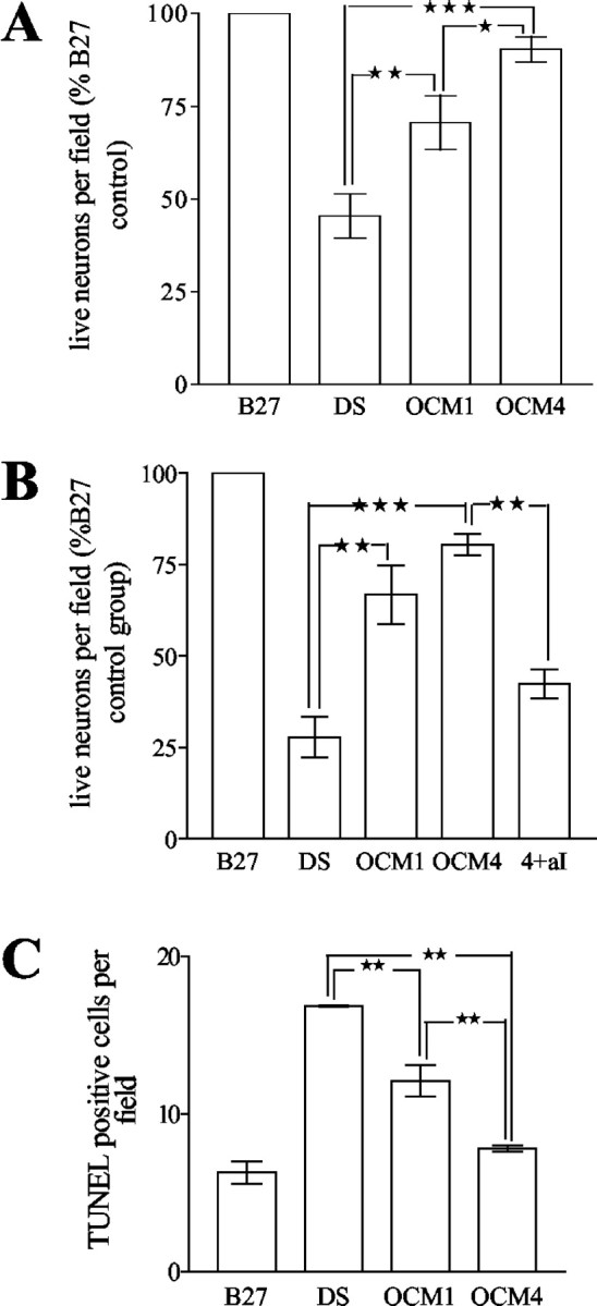
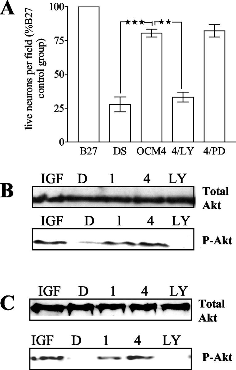
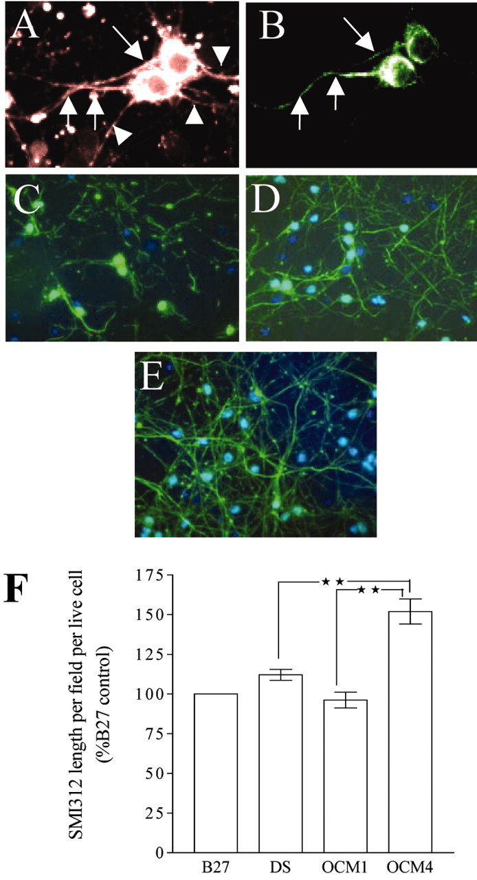
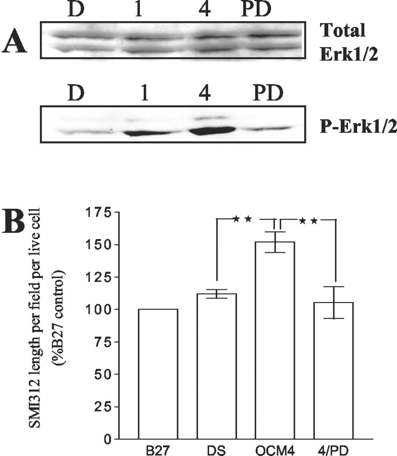
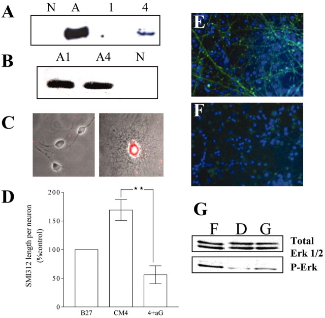
Interactions of CNS cells lead to the establishment of complex neural systems. Specifically, oligodendrocytes form myelin sheaths around axons that enable rapid electrical conduction of impulses. Recent evidence has emerged that oligodendrocytes may also release trophic factors promoting neuronal survival. We therefore studied the effects of factors released from cells of the oligodendrocyte lineage on neuronal survival and also on the morphology of neurons. Neurons derived from rat embryonic cortices were cultured and exposed to media conditioned by oligodendrocyte precursor cells (OPCs) or differentiated oligodendrocytes. In line with previous studies, exposure of OPC and oligodendrocyte-conditioned media (OCM) increased survival, a phosphatidylinositol 3'-kinase (PI3kinase)/Akt-dependent phenomenon. In addition, exposure of neurons to OCM but not OPC conditioned media resulted in increased axonal length per neuron, as detected by antibodies to phosphorylated neurofilaments. OCM exposure resulted in activation of the MAPkinase/extracellular signal-regulated kinase pathway, inhibition of which significantly reduced oligodendrocyte-mediated enhancement of axonal length but, unlike PI3kinase inhibition, had no effect on neuronal survival. Furthermore, we identify glial cell line-derived neurotrophic factor (GDNF) production by differentiated oligodendrocytes and provide evidence that implicates GDNF in OCM-mediated axonal effects, independent of its effect on neuronal survival. Therefore, we have shown that factors released by OPCs and oligodendrocytes induce the activation of distinct intracellular pathways within neurons, which have different functional effects on the cell.


Figures

Figure 1.
Figure 1.
A, Oligodendrocyte-conditioned media increase the survival of young neurons. Effect of nonconditioned medium (DS), medium conditioned by freshly plated OPCs (OCM1), and medium conditioned by predominantly GalC-positive oligodendrocytes (OCM4) on numbers of live (β-tubulin-positive) neurons is shown. Neurons were cultured for 24 hr and exposed to test conditions for 72 hr (n = 5; *p <0.05; **p <0.01; ***p <0.001). B, Oligodendrocyte-conditioned media increase the survival of aged neurons. Effect of nonconditioned medium (DS), OCM1, OCM4, and OCM4 plus neutralizing antibodies to IGF-1 (20 μg/ml; 4 + aI) on numbers of live (β-tubulin-positive) neurons is shown. Neurons were cultured for 120 hr and exposed to test conditions for 72 hr (n = 4; **p < 0.01; ***p < 0.001). C, Oligodendrocyte-conditioned media decrease apoptosis of young neurons. Effect of nonconditioned medium (DS), OCM1, and OCM4 on numbers of TUNEL-positive neurons is shown. Neurons were cultured for 24 hr and exposed to test conditions for 72 hr (n = 3; **p < 0.01).
Figure 2.
Figure 2.
A, PI3kinase inhibitor, but not MAPK/Erk pathway inhibitor, abrogates the OCM4 survival effects on neurons. Effect of nonconditioned medium (DS), OCM4, OCM4 plus LY294002 (10 μm; 4/LY), and OCM4 plus PD98059 (30 μm; 4/PD) on numbers of live (β-tubulin-positive) neurons is shown. Neurons were cultured for 120 hr and exposed to test conditions for 72 hr (n = 4; **p < 0.01; ***p < 0.001). B, C, Oligodendrocyte-conditioned media increase Akt phosphorylation in neurons. B, Akt phosphorylation in young neurons (cultured for 24 hr) exposed to IGF-1 (100 ng/ml; IGF), nonconditioned media (D), OCM1 (1), OCM4 (4), or OCM4 plus LY294002 (10 μm; LY) for 30 min. Bottom, Serine473-phospho-Akt immunoblot. Top, Total-Akt immunoblot of the same membrane. Representative blot of four independent experiments. C, Akt phosphorylation in aged neurons (cultured for 120 hr) and exposed to IGF-1 (100 ng/ml; IGF),nonconditioned media (D), OCM1(1), OCM4(4), or OCM4 plus LY294002 (10 μm; LY) for 30 min. Bottom, Serine473-phospho-Akt immunoblot. Top, Total-Akt immunoblot of the same membrane. Representative blot of three independent experiments.
Figure 3.
Figure 3.
OCM4 increases SMI312-positive process length per cell in aged neurons. Immunographs show neuronal cultures (8 DIV) stained forβ-tubulin (3A) and SMI312 (3B), showing axons (arrows) and other neurites (arrowheads). CE, Effect of nonconditioned medium(C); medium conditioned by freshly plated OPCs (OCM1) (D), and medium conditioned by predominantly GalC-positive oligodendrocytes (OCM4) on SMI312 (phosphorylated neurofilament; green) staining in aged neuronal cultures (E). Nuclei are stained with Hoechst (blue). F, Effect of nonconditioned medium (DS), OCM1, and OCM4 on length of SMI312-stained processes per live neuron in aged neurons (cultured for 120 hr and exposed to test conditions for 72 hr; expressed as a percentage of values obtained from culturing neurons in B27 throughout) (n = 4; **p < 0.01).
Figure 4.
Figure 4.
A, Oligodendrocyte conditioned media increase MAPK/Erk1/2 phosphorylation in aged neurons. MAPK/Erk1/2 phosphorylation in aged neurons (cultured for 120 hr) and exposed to nonconditioned media (D), OCM1 (1), OCM4 (4), and OCM4 plus PD98059 (30 μm; PD) for 30 min is shown. Bottom, Phospho-Erk1/2 immunoblot. Top, Total-Erk1/2 immunoblot of the same membrane. Representative blot of three independent experiments. B, Effect of nonconditioned medium (DS), OCM4, and OCM4 plus PD98059 (4/PD; 30 μm) on the length of SMI312-stained processes per live neuron in aged neurons (cultured for 120 hr and exposed to test conditions for 72 hr; expressed as a percentage of values obtained from culturing neurons in B27 through out) (n = 4; **p < 0.01).
Figure 5.
Figure 5.
A, Oligodendrocyte-conditioned medium contains GDNF. Immunoblots of nonconditioned medium (N) are shown; astrocyte-conditioned medium (A), OCM1(1), and OCM4 (4) are blotted for antibodies against GDNF (representative blots of three independent experiments).B, GDNF production by astrocytes is not influenced by the maturation state of astrocytes. Immunoblots of medium conditioned by highly purified astrocytes were cultured for 1 DIV (A1) and 4 DIV (A4), blotted for antibodies against GDNF and showing comparable levels of GDNF are shown (equal amounts of protein loaded per lane; see Materials and Methods; representative blots of three independent experiments). C, GDNF staining within a differentiated oligodendrocyte. Immunocytochemical staining for GDNF within OPCs (left) and a differentiated oligodendrocyte (right) are shown. Phase contrast picture is shown with superimposed rhodamine-labeled GDNF staining of the same field, showing that differentiated oligodendrocytes contain GDNF. DF, Neutralizing antibodies to GDNF reduce SMI312-positive process length per cell in neurons exposed to OCM4. D, Graph showing effect of OCM4 and OCM4 plus neutralizing antibodies to GDNF (20 μg/ml;4+aG) on length of SMI312-stained processes per live neuron in aged neurons (cultured for 120 hr and exposed to test conditions for 72 hr; expressed as a percentage of values obtained culturing neurons in B27 throughout) (n = 6; **p < 0.01). E, F, Representative immunomicrograph showing the effect of OCM4 (E) and OCM4 plus neutralizing antibodies (F) to GDNF (20 μg/ml) on SMI312 (phosphorylated neurofilament; green) staining in aged neuronal cultures. Nuclei stained with Hoechst are shown (blue). G, GDNF increases MAPK/Erk1/2 phosphorylation in aged neurons. Immunoblot showing MAPK/Erk1/2 phosphorylation in aged neurons (cultured for 120 hr) and exposed to DMEM/10% fetal calf serum (F), nonconditioned media (D), and nonconditioned media plus recombinant GDNF (10 ng/ml; G) for 30 min. Bottom panel, Phospho-Erk1/2 immunoblot. Top panel, Total-Erk1/2 immunoblot of the same membrane (representative immunoblot of four independent experiments).

References

    1. Acheson A, Conover JC, Fandl JP, DeChiara TM, Russell M, Thadani A, Squinto SP, Yancopoulos GD, Lindsay RM ( 1995) A BDNF autocrine loop in adult sensory neurons prevents cell death. Nature 374: 450–453. - PubMed
    1. Arce V, Pollock RA, Philippe JM, Pennica D, Henderson CE, deLapeyriere O ( 1998) Synergistic effects of schwann- and muscle-derived factors on motoneuron survival involve GDNF and cardiotrophin-1 (CT-1). J Neurosci 18: 1440–1448. - PMC - PubMed
    1. Atwal JK, Massie B, Miller FD, Kaplan DR ( 2000) The TrkB-Shc site signals neuronal survival and local axon growth via MAPK/Erk pathway and PI3-kinase. Neuron 27: 265–277. - PubMed
    1. Banker GA ( 1980) Trophic interactions between astroglial cells and hippocampal neurons in culture. Science 209: 809–810. - PubMed
    1. Brady ST, Witt AS, Kirkpatrick LL, de Waegh SM, Readhead C, Tu PH, Lee VM ( 1999) Formation of compact myelin is required for the maturation of the axonal cytoskeleton. J Neurosci 19: 7278–7288. - PMC - PubMed

Publication types

MeSH terms

Substances

LinkOut - more resources