. 2003 Jun 15;23(12):5105-12.
doi: 10.1523/JNEUROSCI.23-12-05105.2003.

Factors in the genetic background suppress the engrailed-1 cerebellar phenotype

Affiliations

Factors in the genetic background suppress the engrailed-1 cerebellar phenotype

Natalie A Bilovocky et al. J Neurosci. .

Abstract

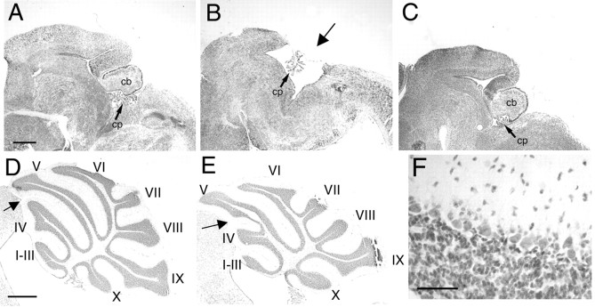
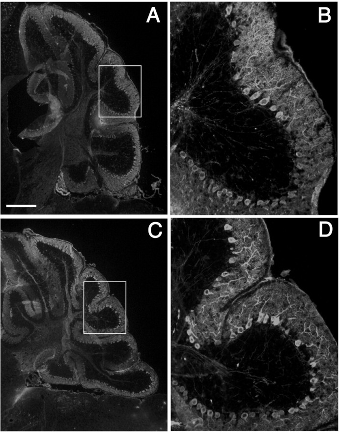
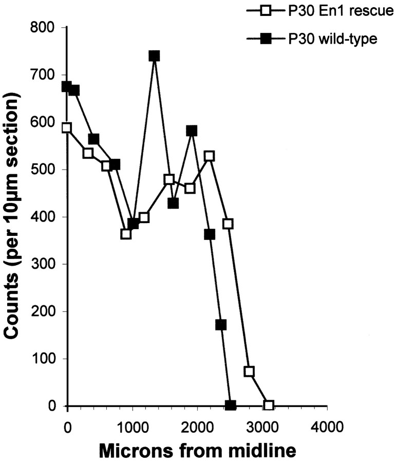
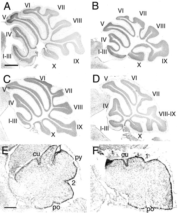
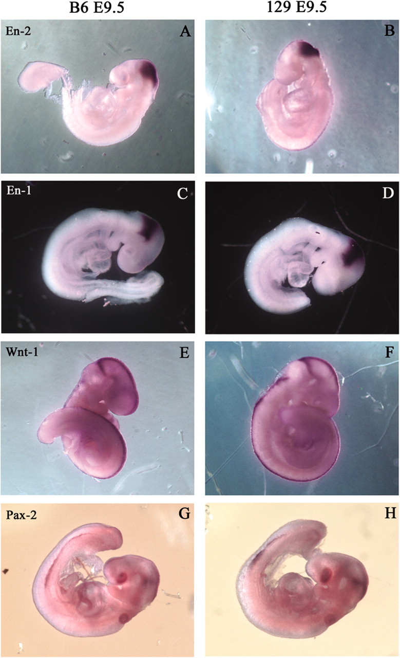
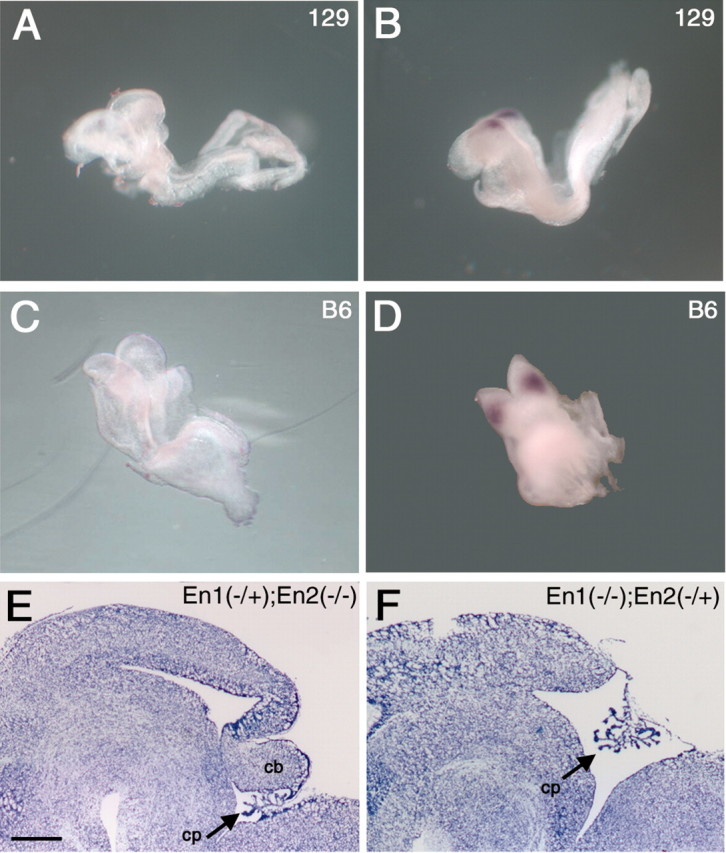
The mouse homeodomain protein, Engrailed-1, is generally viewed as an essential player in the early establishment and maintenance of the midbrain/hindbrain region that gives rise to the cerebellum and midbrain. In keeping with this, engineered null mutations at this locus have been reported to lead to perinatal lethality accompanied by near-total absence of cerebellar and caudal midbrain structures. We report here that these cerebellar phenotypes are nearly completely suppressed on a C57BL/6J genetic background. All cell types are present and arranged properly in both the cortex and the deep nuclei, and cell counts reveal no significant absence of cerebellar Purkinje cells. Folial patterns are nearly normal, although an apparent fusion of lobules IV and V is consistently noted. Significantly, no change in the Engrailed-2 mutant phenotype occurs after a similar background switch, and whole-mount in situ hybridization reveals identical En2 expression patterns in wild-type C57BL/6J and 129/Sv mice. One likely mechanism for the En1-/- phenotype suppression is a temporal and/or spatial change in the pattern of Engrailed-2 expression apparent only in the absence of Engrailed-1. In support of this, C57BL/6-En1-/- embryos that are also En2+/- lack a cerebellum and caudal midbrain: a phenotype identical to 129/Sv-En1-/- mice.


Figures

Figure 1.
Figure 1.
Cerebellar phenotype of En1Hd/Hd mutants on 129/Sv and C57BL/6J backgrounds. Cresyl violet-stained sagittal cryostat sections of E17.5 midbrain/hindbrain region of C57BL/6J wild-type (A), 129/Sv—En1Hd/Hd (B), and C57BL/6J—En1Hd/Hd (C). The arrow in B indicates where the cerebellum would normally appear in the 129/Sv mutant. Comparison between adult C57BL/6J wild-type cerebellum (D) and C57BL/6J—En1Hd/Hd rescue cerebellum (E) reveals a partial fusion of vermal lobes IV and V, but no other abnormalities. At higher magnification (F), the microarchitecture of the cerebellar cortex is indistinguishable from normal. cb, Cerebellum; cp, choroid plexus. Scale bars: (in D), A—E, 500 μm; F, 25 μm.
Figure 2.
Figure 2.
Purkinje cell morphology in adult C57BL/6J—En1Hd/Hd cerebella are indistinguishable from normal. Sagittal cryostat sections immunostained for calbindin, a Purkinje cell-specific calcium-binding protein; the C57BL/6J wild-type [A, B (magnification of inset in A)] and C57BL/6J—En1Hd/Hd cerebella [C, D (magnification of inset in C)] show no difference in either the density of Purkinje cell bodies or the complexity of their dendritic arborizations. Scale bar: (in A) A, C, 400 μm; B, D, 100 μm.
Figure 3.
Figure 3.
No detectable Purkinje cell loss occurs in the cerebella of rescue animals. Two P30 C57BL/6J animals of each genotype (En1+/+ and En1Hd/Hd) were analyzed. There is no significant difference in Purkinje cell number between the two genotypes (p = 0.29; one-tailed Student's t test). The raw counts per 10 μm section plotted as a function of distance from the midline is shown.
Figure 4.
Figure 4.
Mutant limb abnormalities are not suppressed in C57BL/6J—En1Hd/Hd animals; they are identical to those seen on the 129/Sv background. A—F, Adult paws of C57BL/6J wild-type (A, C, E) and C57BL/6J—En1Hd/Hd (B, D, F) animals. Forelimbs are shown in A, B, E, and F; hindlimbs in C and D. Arrows in B denote a sixth postaxial digit (left arrow) and a fusion of digits I and II (right arrow). Ectopic footpads and hair growth are also found on the ventral surface of rescue limbs (B). The right hindlimb of a rescue mutant (D) showed similar abnormalities, but generally hindlimb defects were less severe. The nails of the wild-type digits (E) are hooked, with a clear dorsal (d)/ventral (v) asymmetry. In contrast, the nails of the C57BL/6J—En1Hd/Hd paw (F) are symmetrical and straight.
Figure 5.
Figure 5.
Strain background does not alter the En2Hd/Hd phenotype. Cresyl violet-stained sagittal paraffin sections of 129/Sv—En2Hd/Hd adults (B) revealed a posterior lobe defect in which the VIIIth and IXth vermal lobes are fused, unlike in the 129/Sv wild-type (A). Sagittal sections of C57BL/6J—En2Hd/Hd adults (D) display abnormal vermal lobe branching, similar to that of the 129/Sv—En2Hd/Hd mutants, but quite different from the branching seen in the C57BL/6J wild-type (C). Cresyl violet-stained sagittal cryostat sections of newborn C57BL/6J—En2Hd/Hd mice (F) reveal abnormal fissure formation in the posterior region of the cerebellum compared with wild-type C57BL/6J (E). The secondary (2) and prepyramidal (py) fissures are less obvious in the developing mutants, whereas the anterior fissures appear normal. cu, Preculminate; 1, primary; po, posterolateral. Scale bars: (in A) A—D, 500 μm; (in E) E, F, 200 μm.
Figure 6.
Figure 6.
Expression patterns of early pattern formation genes are not affected by strain background. Whole-mount in situ hybridization of E9.5 embryos for En2 (A, B), En1 (C, D), Wnt1 (E, F), and Pax2 (G, H). Two different strains were used, C57BL/6J (A, C, E, G) and 129/Sv (B, D, F, H). No differences are seen between the two strains.
Figure 7.
Figure 7.
There is no difference in onset of En2 expression between 129/Sv (A, B) and C57BL/6J (C, D) wild-type mice. Whole-mount in situ hybridization of the 4- to 5-somite stage (A, C) shows no expression in either strain, whereas at the 6-somite stage (B, D), expression is first detected. Despite this, in the absence of En1, development of the cerebellum depends critically on En2 expression. Cresyl violet-stained sagittal cryostat sections of E17.5 (En1Hd/Hd; En2Hd/+) mutants (F) reveal a lack of cerebellum, whereas the(En1Hd/+; En2Hd/Hd) mutants (E) show no such loss. Both E and F are on a C57BL/6J background. Scale bar: (in E) E, F, 500 μm.

References

    1. Bonyadi M, Rusholme SA, Cousins FM, Su HC, Biron CA, Farrall M, Akhurst RJ ( 1997) Mapping of a major genetic modifier of embryonic lethality in TGF β1 knockout mice. Nat Genet 15: 207–211. - PubMed
    1. Davis C, Joyner A ( 1988) Expression patterns of the homeobox-containing genes En-1 and En-2 and the proto-oncogene int-1 diverge during mouse development. Genes Dev 2: 1736–1744. - PubMed
    1. Davis C, Noble-Topham S, Rossant J, Joyner A ( 1988) Expression of the homeobox-containing gene En-2 delineates a specific region of the developing mouse brain. Genes Dev 2: 361–371. - PubMed
    1. Gerlai R, Millen KJ, Herrup K, Fabien K, Joyner AL, Roder J ( 1996) Impaired motor learning performance in cerebellar En-2 mutant mice. Behav Neurosci 110: 126–133. - PubMed
    1. Greferath U, Bennie A, Kourakis A, Bartlett PF, Murphy M, Barrett GL ( 2000) Enlarged cholinergic forebrain neurons and improved spatial learning in p75 knockout mice. Eur J Neurosci 12: 885–893. - PubMed

Publication types

MeSH terms