. 2003 Jun 15;23(12):5131-40.
doi: 10.1523/JNEUROSCI.23-12-05131.2003.

Human embryonic germ cell derivatives facilitate motor recovery of rats with diffuse motor neuron injury

Affiliations

Human embryonic germ cell derivatives facilitate motor recovery of rats with diffuse motor neuron injury

Douglas A Kerr et al. J Neurosci. .

Abstract

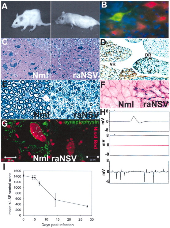
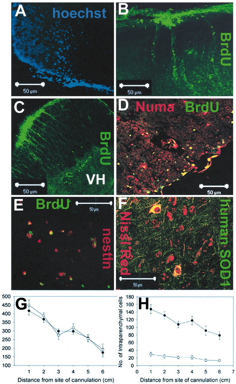
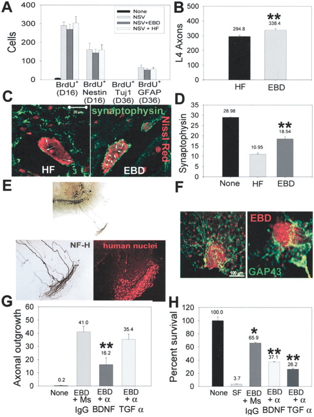
We have investigated the potential of human pluripotent cells to restore function in rats paralyzed with a virus-induced motor neuronopathy. Cells derived from embryonic germ cells, termed embryoid body-derived (EBD) cells, introduced into the CSF were distributed extensively over the rostrocaudal length of the spinal cord and migrated into the spinal cord parenchyma in paralyzed, but not uninjured, animals. Some of the transplanted human cells expressed the neuroglial progenitor marker nestin, whereas others expressed immunohistochemical markers characteristic of astrocytes or mature neurons. Rare transplanted cells developed immunoreactivity to choline acetyltransferase (ChAT) and sent axons into the sciatic nerve as detected by retrograde labeling. Paralyzed animals transplanted with EBD cells partially recovered motor function 12 and 24 weeks after transplantation, whereas control animals remained paralyzed. Semi-quantitative analysis revealed that the efficiency of neuronal differentiation and extension of neurites could not account for the functional recovery. Rather, transplanted EBD cells protected host neurons from death and facilitated reafferentation of motor neuron cell bodies. In vitro, EBD cells secrete transforming growth factor-alpha (TGF-alpha) and brain-derived neurotrophic factor (BDNF). Neutralizing antibodies to TGF-alpha and to BDNF abrogated the ability of EBD-conditioned media to sustain motor neuron survival in culture, whereas neutralizing antibodies to BDNF eliminated the axonal outgrowth from spinal organotypics observed with direct coculture of EBD cells. We conclude that cells derived from human pluripotent stem cells have the capacity to restore neurologic function in animals with diffuse motor neuron disease via enhancement of host neuron survival and function.


Figures

Figure 1.
Figure 1.
Neuroadapted Sindbis virus causes progressive lower motor neuron injury in rats. A, Progressive kyphoscoliosis (left) and then hindlimb paralysis (right) develop in rats after intracranial infection of 1000 PFU of rat-adapted Sindbis virus (raNSV). B—E, Effect of raNSV on motor neurons. Rats were given intracranial infections of NSV encoding green fluorescent protein (raNSVGFP). At 3 d later the spinal cord sections were costained with Nissl red and Hoechst 33342. B, Note the prominent green fluorescence in spinal motor neurons. C, Lumbar enlargement of spinal cord 28 d after raNSV infection. Note a significant reduction in motor neurons in paralyzed rat (right) as compared with nonparalyzed rat (left). D, Silver-stained sections of lumbar spinal cord 28 d after raNSV infection. Note the selective depletion of axons in the ventral root (VR) in a paralyzed rat and preservation of dorsal roots(DR).E, Toluidine blue-stained sections (1 μm) of the L4 ventral root from uninfected rat (left) and 28 d after infection (right) show prominent Wallerian degeneration. F, Muscle biopsy of paralyzed (right) and control (left) rat. Note neurogenic atrophy with angulated and degenerating fibers in the paralyzed rat, with marked variation in fiber size. G, Loss of presynaptic input to surviving motor neurons after raNSV infection. Note the reduction in synaptophysin contacts (green; arrows reflect synaptic contacts) onto motor neurons 28 d after infection (right) as compared with uninfected motor neurons. H, Electrophysiologic recordings of nonparalyzed and paralyzed rats. Shown are normal compound motor unit potentials in nonparalyzed rat (5.12 mV; top), absent motor action potential in severely paralyzed rat (middle), and positive sharp waves on EMG in raNSV-paralyzed rat (bottom). I, Temporal course of motor neuron loss after raNSV infection as defined by quantitation of ventral L4 root axons. Stereologic examination of semithin sections was performed. Sections from three animals were examined at each time point. Data are presented as the mean at each time point ± SE, with the y-axis representing number of ventral axons.
Figure 2.
Figure 2.
EBD cells introduced into the lumbar CSF migrate into the spinal cord and widely distribute over the length of the spinal cord. A—F, Histologic analysis of rats 4 weeks after implantation revealed intraparenchymal engraftment of EBD cells with multiple distinct markers. Cells prelabeled with Hoechst (A) or BrdU (B—E) showed migration into the spinal cord from the subarachnoid space. Immunohistochemical analysis of spinal cords with antisera specific to human Numa (D) or human SOD1 (F) confirmed migration into the spinal cord. G, Quantitation of the rostrocaudal distribution of all surviving cells (meningeal and intraspinal) at 4 weeks after implantation relative to the cannula placement. Every 50th axial spinal cord section was examined for the number of cells that exhibited human nuclear matrix antigen positivity. Five nonparalyzed EBD-transplanted animals and five paralyzed EBD-transplanted animals were analyzed. EBD cells were detected at all levels of the neuraxis; EBD cell survival was similar in raNSV-paralyzed (filled circles) and in nonparalyzed animals (open circles). H, When only intraparenchymal EBD cells (Numa +) were counted, a greater migration of cells into the spinal cord was seen in paralyzed animals (filled circles) as compared with nonparalyzed (open circles) animals (p = 0.001). In both graphs, data are presented at each distance from the cannulation as mean ± SE.
Figure 3.
Figure 3.
Some transplanted EBD cells acquire immunohistochemical markers of neural cells. At 28 d after implantation spinal cord sections were generated and examined by immunohistochemistry to determine whether the implanted EBD cells expressed neuronal markers such as GFAP (A), β3-tubulin (B), MAP2 (C), or ChAT (D). Orthogonal images confirm the colocalization of GFAP, β3-tubulin, and ChAT with human-specific nuclear matrix antigen staining. E,F, Rare retrograde labeling of prelabeled EBD cells from the sciatic nerve. E, Rare Hoechst-prelabeled EBD cells (blue) in the lumbar gray matter that were retrogradely labeled from the sciatic nerve (green) and were strongly Nissl+ (red). Retrograde labeling (green) of EBD cells in the field (top left) reveals two large and one smaller cell in the ventral gray matter. Nissl red staining (top right) reveals four Nissl+ cells in the field, including the three that were retrogradely labeled. Hoechst staining (bottomleft) reveals that one of the retrogradely labeled Nissl+ cells is EBD in origin, whereas several additional EBD cells were not retrogradely labeled and one additional EBD cell acquired strong Nissl+ staining (but did not retrogradely label). The asterisk denotes an EBD-derived cell that retrogradely labeled the sciatic nerve. F, Numa+ EBD cells (red) that retrogradely label from the sciatic nerve (green) with orthogonal views confirming colocalization. Note the nonretrogradely labeled EBD cell (red only, top right) and the host cell that retrogradely labeled (green only). Control animals that had a second sciatic nerve transection more proximal to the original site to prevent axonal transport of the chemical never exhibited any intraspinal label.
Figure 5.
Figure 5.
EBD transplantation facilitates host motor neuron survival and axonal reafferentation of host motor neurons. A, EBD transplantation did not increase the proliferation or differentiation of endogenous spinal neural stem cells. Rats were given BrdU for 3d from days 12–15 after infection and then were killed at either day 16 or day 36 after infection. Immunohistochemical analysis determined the frequency of BrdU+ cells per section or the colocalization of BrdU with nestin, β-tubulin, or GFAP. Although raNSV induced neural stem cell proliferation within the spinal cord, no new neurons could be detected at day 36 with any of the transplantation paradigms. B, Quantitation of the number of L4 ventral root axons at 6 months after transplantation shows a significant increase in surviving axons in transplanted animals as compared with control-transplanted animals. L4 nerve roots from 15 animals in each group were analyzed; the comparisons were made with a Student's t test (p < 0.03). C, Representative photomicrographs of the synaptophysin complement on motor neurons at 6 months after transplantation in HF-transplanted (left) and EBD-transplanted (right) rats. D, The number of synaptophysin contacts per motor neuron cell body was enhanced in transplanted animals at 6 months: 50 motor neurons were counted in each of five animals per group. Motor neurons in HF-transplanted rats had 10.95 ± 0.65 synaptophysin contacts per soma, whereas EBD-transplanted rats had 18.54 ± 1.14 synaptophysin contacts per soma. Comparisons between HF- and EBD-transplanted animals were performed by the Mann—Whitney test (p < 0.02). E, F, EBD cells exhibit tropic properties for host axons in a spinal cord organotypic model. EBD cells were placed outside the organotypic margin and cultured for an additional 2 weeks. E, In the presence of EBD cells the host axons were stimulated to send out processes from the organotypic margin for up to 1.2 mm after 14 d. The bottom panels represent a higher power view of axonal growth (left) into the cluster of human nuclei-labeled EBD cells (right). F, Stimulation of GAP 43 expression (green) by host axons in response to EBD cells (red). G, Inhibition of EBD-stimulated axonal growth in organotypic cultures by neutralizing antibodies to BDNF, but not TGF-α. Spinal cord organotypic cultures were cocultured with EBD cells as above, with neutralizing antibodies to BDNF, TGF-α, or control antibody. The scores are depicted as the number of Smi32+ axons that have grown out of the margin of the organotypic culture per slice (p = 0.007). H, Conditioned medium from EBD cells allowed for survival of cultured motor neurons in the absence of exogenous growth factors (compare serum-free medium, lane two, with EBD-conditioned medium, lane 3) (*p = 0.001 compared with lane 2). The addition of neutralizing antibodies to either BDNF or TGF-α partially abolished the ability of EBD-conditioned medium to support motor neuron survival. **p < 0.001 compared with lane 3.
Figure 4.
Figure 4.
Functional recovery of EBD-transplanted rats at various intervals. In all, 15 paralyzed rats in each group were implanted with 300,000 EBD cells, baby hamster kidney (BHK) cells, or human fibroblast (HF) cells and were scored for recovery by two independent raters who were blinded to the treatment groups. A, BHK-transplanted (open triangles) and HF-transplanted (filled circles) rats exhibited no recovery of locomotor function at 4, 8, 12, or 24 weeks. EBD-transplanted rats showed a significant functional improvement as compared with BHK- or HF-transplanted animals at 12 and 24 weeks. Asterisks denote significant recovery of EBD-transplanted animals (p = 0.004 at 12 weeks; p = 0.0001 at 24 weeks). B, Measurement of hindlimb strength via a digital force gauge revealed improved grip strength in transplanted animals (filled triangles), whereas HF-transplanted (filled circles) and BHK-transplanted (open triangles) animals did not. By 6 months after transplantation the EBD-transplanted animals had recovered ∼40% of their pre-paralysis strength, whereas there had been no recovery in the other groups. Asterisks denote significant improvement in hindlimb strength (p < 0.001 at 12 and 24 weeks). C, Measurement of righting time as an independent and objective measure of motor recovery. All 15 animals transplanted with either EBD or HF cells were tested for the time required to right themselves when placed in a supine position. Two separate trials were conducted, and the total time required for both trials was summed at the time of transplantation and again at 24 weeks after transplantation. The data are presented as a Δ score, reflecting an individual animal's change from its baseline. Positive numbers reflect improved performance on this task, whereas negative numbers reflect worse performance. In all, 11 of 15 EBD-transplanted animals (back row) had an improvement of ≥3 sec, whereas only 1 of 15 HF-transplanted animals did.

Similar articles

Cited by

References

    1. Barde YA, Davies AM, Johnson JE, Lindsay RM, Thoenen H ( 1987) Brain-derived neurotrophic factor. Prog Brain Res 71: 185–189. - PubMed
    1. Basso DM, Beattie MS, Bresnahan JC ( 1995) A sensitive and reliable locomotor rating scale for open field testing in rats. J Neurotrauma 12: 1–21. - PubMed
    1. Benedetti S, Pirola B, Pollo B, Magrassi L, Bruzzone MG, Rigamonti D, Galli R, Selleri S, Di Meco F, De Fraja C, Vescovi A, Cattaneo E, Finocchiaro G ( 2000) Gene therapy of experimental brain tumors using neural progenitor cells. Nat Med 6: 447–450. - PubMed
    1. Benraiss A, Chmielnicki E, Lerner K, Roh D, Goldman SA ( 2001) Adenoviral brain-derived neurotrophic factor induces both neostriatal and olfactory neuronal recruitment from endogenous progenitor cells in the adult forebrain. J Neurosci 21: 6718–6731. - PMC - PubMed
    1. Boillee S, Cadusseau J, Coulpier M, Grannec G, Junier MP ( 2001) Transforming growth factor α: a promoter of motoneuron survival of potential biological relevance. J Neurosci 21: 7079–7088. - PMC - PubMed

Publication types

MeSH terms

LinkOut - more resources