. 2003 Jun 15;23(12):5219-26.
doi: 10.1523/JNEUROSCI.23-12-05219.2003.

Selectively reduced expression of synaptic plasticity-related genes in amyloid precursor protein + presenilin-1 transgenic mice

Affiliations

Selectively reduced expression of synaptic plasticity-related genes in amyloid precursor protein + presenilin-1 transgenic mice

Chad A Dickey et al. J Neurosci. .

Abstract

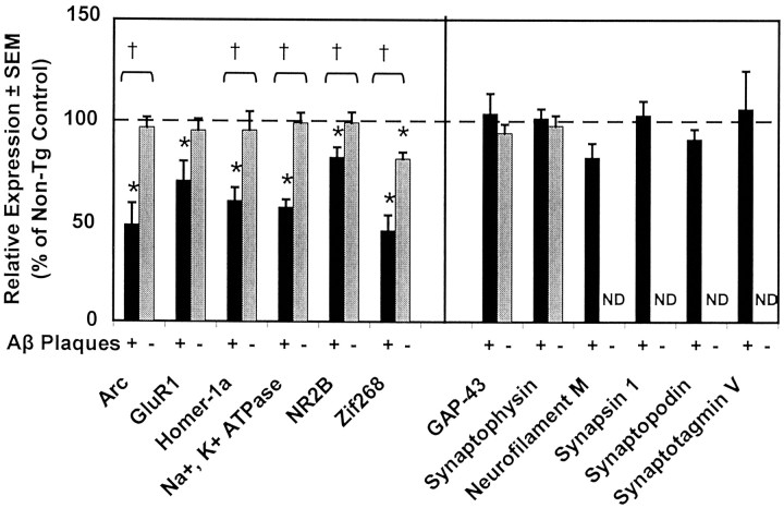
A critical question in Alzheimer's disease (AD) research is the cause of memory loss that leads to dementia. The amyloid precursor protein + presenilin-1 (APP+PS1) transgenic mouse is a model for amyloid deposition, and like AD, the mice develop memory deficits as amyloid deposits accumulate. We profiled gene expression in these transgenic mice by microarray and quantitative RT-PCR (qRT-PCR). At the age when these animals developed cognitive dysfunction, they had reduced mRNA expression of several genes essential for long-term potentiation and memory formation (Arc, Zif268, NR2B, GluR1, Homer-1a, Nur77/TR3). These changes appeared to be related to amyloid deposition, because mRNA expression was unchanged in the regions that did not accumulate amyloid. Transgene expression was similar in both amyloid-containing and amyloid-free regions of the brain. Interestingly, these changes occurred without apparent changes in synaptic structure, because a number of presynaptic marker mRNAs (growth-associated protein-43, synapsin, synaptophysin, synaptopodin, synaptotagmin, syntaxin) remained stable. Additionally, a number of genes related to inflammation were elevated in transgenic mice, primarily in the regions containing amyloid. In AD cortical tissue, the same memory-associated genes were downregulated. However, all synaptic and neuronal transcripts were reduced, implying that the loss of neurons and synapses contributed to these changes. We conclude that reduced expression of selected genes associated with memory consolidation are linked to memory loss in both circumstances. This suggests that the memory loss in APP+PS1 transgenic mice may model the early memory dysfunction in AD before the degeneration of synapses and neurons.


Figures

Figure 1.
Figure 1.
Gene expression profile of transgenic mice in amyloid-containing and amyloid-free brain regions by qRT-PCR. The differential expression (transgenic expression relative to nontransgenic expression) is presented for genes that are downregulated in transgenic animals and primarily postsynaptic (left half of figure) and those that are stably expressed and essentially presynaptic (right half of figure). The relative expression in the amyloid-containing region (hippocampus; black bars) and the amyloid-free region (cerebellum; gray bars) is shown when both regions were analyzed. These regions are also designated in the line to the right of Aβ Plaques with a + symbol beneath the bar indicating amyloid-containing region or a − symbol beneath the bar indicating an amyloid-free region. After qRT-PCR, each gene transcript level for each sample is first normalized to the 18S RNA level measured from the same RT. For each region, relative expression was determined by dividing each transgenic value by the average of the nontransgenic values. The relative expression values for the amyloid-containing and amyloid-free regions could also be compared statistically to determine whether the reduction in expression was different in the two structures. The values represented in this figure are the mean ± SEM. We did not determine cerebellum measurements for several genes that were unaffected in the hippocampus of the transgenics, as indicated by ND in place of the bar on the figure. Asterisks indicate significant differences between APP+PS1 mice and nontransgenic littermates (p < 0.05). † symbol above the bracket indicates transgene-associated downregulation to a greater extent (p < 0.05) in the amyloid-containing than the amyloid-free regions.
Figure 2.
Figure 2.
Gene expression profile of Alzheimer disease tissue in amyloid-containing and amyloid-free brain regions by qRT-PCR. The differential expression (Alzheimer relative to age-matched normals) is presented for genes that are primarily postsynaptic (left half of figure) and those that are primarily presynaptic (right half of figure). The relative expression in the amyloid-containing region (temporal cortex; area 21; black bars) and amyloid-free region (cerebellum; gray bars) is shown when both regions were analyzed. These regions are also designated in the line to the right of Aβ Plaques with a + symbol beneath the bar indicating amyloid containing region or a − symbol beneath the bar indicating an amyloid-free region. After qRT-PCR, each gene transcript level for each sample is first normalized to 18S RNA measurements from the same RT. For each region, relative expression was determined by dividing each Alzheimer sample value by the average of the age-matched normal values. The relative expression values for the amyloid-containing and amyloid-free regions could also be compared statistically to determine whether the reduction in expression was different in the two structures. The values represented in this figure are the mean ± SEM. We did not determine cerebellum measurements for several genes as indicated by ND in place of the bar on the figure. Asterisks indicate significant differences between Alzheimer and age-matched normals (p < 0.05). † symbol above the bracket indicates disease-associated downregulation to a greater extent (p < 0.05) in the amyloid-containing than the amyloid-free regions.

References

    1. Ango F, Pin JP, Tu JC, Xiao B, Worley PF, Bockaert J, Fagni L ( 2000) Dendritic and axonal targeting of type 5 metabotropic glutamate receptor is regulated by homer1 proteins and neuronal excitation. J Neurosci 20: 8710–8716. - PMC - PubMed
    1. Arendash GW, King DL, Gordon MN, Morgan D, Hatcher JM, Hope CE, Diamond DM ( 2001) Progressive, age-related behavioral impairments in transgenic mice carrying both mutant amyloid precursor protein and presenilin-1 transgenes. Brain Res 891: 42–53. - PubMed
    1. Austin LA, Arendash GW, Gordon G, Diamond DM, DiCarlo G, Dickey C, Ugen KE, Morgan D ( 2003) Short-term A-beta vaccinations do not improve cognitive performance in cognitively-impaired APP+PS1 mice. Behav Neurosci, in press. - PubMed
    1. Bi H, Sze CI ( 2002) N-methyl-d-aspartate receptor subunit NR2A and NR2B messenger RNA levels are altered in the hippocampus and entorhinal cortex in Alzheimer's disease. J Neurol Sci 200: 11–18. - PubMed
    1. Bolshakov VY, Golan H, Kandel ER, Siegelbaum SA ( 1997) Recruitment of new sites of synaptic transmission during the cAMP-dependent late phase of LTP at CA3–CA1 synapses in the hippocampus. Neuron 19: 635–651. - PubMed

Publication types

MeSH terms