

Does a betaretrovirus infection trigger primary biliary cirrhosis?
- PMID: 12832623
- PMCID: PMC166250
- DOI: 10.1073/pnas.1433063100
Does a betaretrovirus infection trigger primary biliary cirrhosis?
Abstract
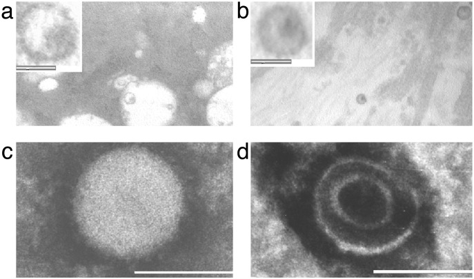
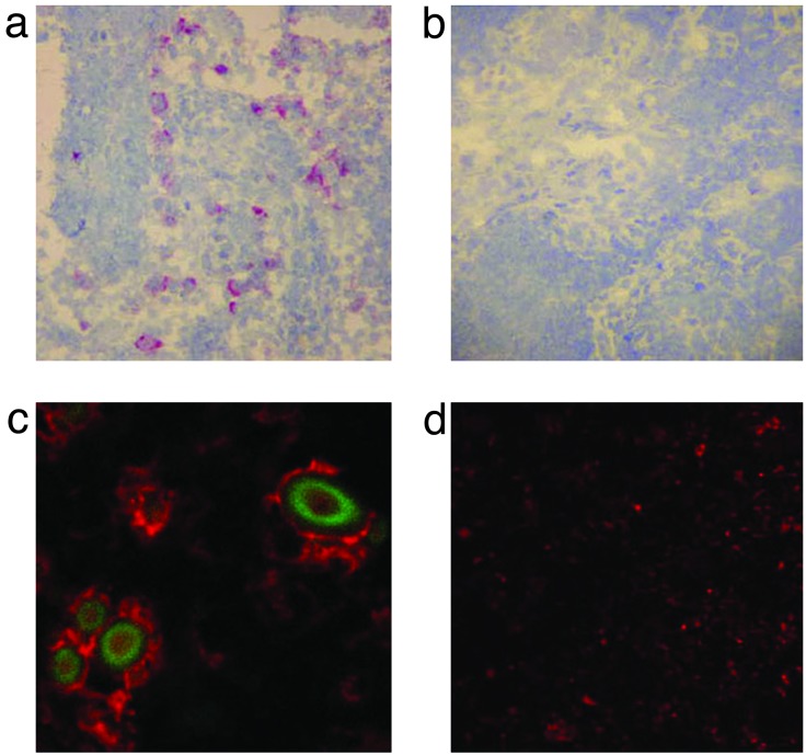
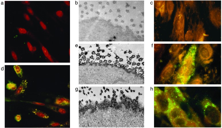
Patients with primary biliary cirrhosis develop progressive ductopenia associated with the production of antimitochondrial antibodies that react with a protein aberrantly expressed on biliary epithelial cells and peri-hepatic lymph nodes. Although no specific microbe has been identified, it is thought that an infectious agent triggers this autoimmune liver disease in genetically predisposed individuals. Previous serologic studies have provided evidence to suggest a viral association with primary biliary cirrhosis. Here we describe the identification of viral particles in biliary epithelium by electron microscopy and the cloning of exogenous retroviral nucleotide sequences from patients with primary biliary cirrhosis. The putative agent is referred to as the human betaretrovirus because it shares close homology with the murine mammary tumor virus and a human retrovirus cloned from breast cancer tissue. In vivo, we have found that the majority of patients with primary biliary cirrhosis have both RT-PCR and immunohistochemistry evidence of human betaretrovirus infection in lymph nodes. Moreover, the viral proteins colocalize to cells demonstrating aberrant autoantigen expression. In vitro, we have found that lymph node homogenates from patients with primary biliary cirrhosis can induce autoantigen expression in normal biliary epithelial cells in coculture. Normal biliary epithelial cells also develop the phenotypic manifestation of primary biliary cirrhosis when cocultivated in serial passage with supernatants containing the human betaretrovirus or the murine mammary tumor virus, providing a model to test Koch's postulates in vitro.
Figures
References
-
- Neuberger, J. (1997) Lancet 350 875-879. - PubMed
-
- Nishio, A., Keeffe, E. B. & Gershwin, M. E. (2002) Semin. Liver Dis. 22 211-314. - PubMed
-
- Joplin, R. & Gershwin, M. E. (1997) Semin. Liver Dis. 17 97-103. - PubMed
-
- Harada, K., Van de Water, J., Leung, P. S., Coppel, R. L., Nakanuma, Y. & Gershwin, M. E. (1997) Hepatology 25 27-32. - PubMed
Publication types
MeSH terms
Substances
Associated data
- Actions
- Actions
- Actions
- Actions
- Actions
- Actions
- Actions
- Actions
- Actions
- Actions
- Actions
- Actions
- Actions
- Actions
- Actions
- Actions
- Actions
- Actions
- Actions
Grants and funding
LinkOut - more resources
Full Text Sources
Other Literature Sources
Medical
