Review
. 2003 Jul;56(7):497-506.
doi: 10.1136/jcp.56.7.497.

Salivary gland-like tumours of the breast: surgical and molecular pathology

Affiliations
Review

Salivary gland-like tumours of the breast: surgical and molecular pathology

M Pia-Foschini et al. J Clin Pathol. 2003 Jul.

Erratum in

  • J Clin Pathol. 2003 Oct;56(10):804

Abstract

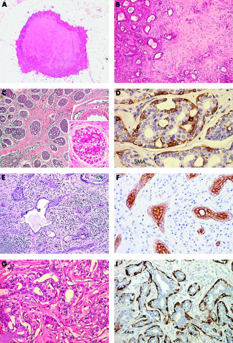
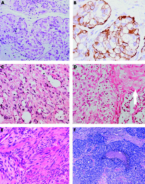
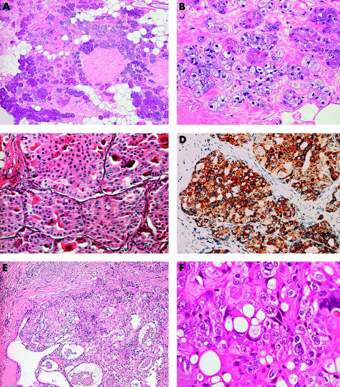
Breast glands and salivary glands are tubulo-acinar exocrine glands that can manifest as tumours with similar morphological features, but that differ in incidence and clinical behaviour depending on whether they are primary in breast or salivary glands. Salivary gland-like tumours of the breast are of two types: tumours with myoepithelial differentiation and those devoid of myoepithelial differentiation. The first and more numerous group comprises a spectrum of lesions ranging from "bona fide" benign (such as benign myoepithelioma and pleomorphic adenoma), to low grade malignant (such as adenoid cystic carcinoma, low grade adenosquamous carcinoma, and adenomyoepithelioma), to high grade malignant lesions (malignant myoepithelioma). The second group comprises lesions that have only recently been recognised, such as acinic cell carcinoma, oncocytic carcinoma of the breast, and the rare mucoepidermoid carcinoma.


Figures

Figure 1
Figure 1
Benign/low grade malignant salivary gland-like tumours with myoepithelial differentiation. (A) Mammary pleomorphic adenoma has circumscribed margins and shows a central area of cartilaginous tissue; (B) medium power magnification disclosing glandular structures immersed in a myxoid stroma. (C) Adenoid cystic carcinoma (ACC) of the breast showing solid and cribriform architecture; inset, breast ACC is composed of basaloid cells that outline spaces containing basal-like material and of eosinophilic cells lining true glandular lumina; (D) staining for smooth muscle actin (SMA) demonstrates myoepithelial cells. (E) Syringomatous carcinoma/low grade adenosquamous carcinoma (SyC/LGASC) is composed of angulated glands located in desmoplastic stroma; (F) SyC squamous differentiation is frequent and contains high molecular weight keratin. (G) Breast adenomyoepithelioma (AME): glandular structures are formed by an inner layer of epithelial cells with eosinophilic cytoplasm and by an outer layer of myoepithelial cells with clear cytoplasm; (H) staining for calponin demonstrates myoepithelial cells in breast AME.
Figure 2
Figure 2
Malignant myoepithelioma/myopithelial carcinoma. (A) Malignant myoepithelioma (MM) with intralobular growth; (B) myoepithelial differentiation is confirmed by anti-smooth muscle actin antibody; (C) MM is composed of a solid proliferation of extremely atypical cells; (D) foci of squamous differentiation are occasionally seen (arrow); (E) MM with spindle cell morphology (note the high mitotic activity). (F) Grade III invasive ductal carcinoma with myoepithelial differentiation.
Figure 3
Figure 3
Salivary gland-like tumours without myoepithelial differentiation. (A) Acinic cell carcinoma (AcCC) with microglandular growth; (B) cells have a granular eosinophilic to amphophilic cytoplasm; some cells also show foamy cytoplasm (courtesy of Dr IO Ellis). (C) Breast oncocytic carcinoma (OC) is composed of cells with granular eosinophilic cytoplasm; (D) anti-mitochondrion antibody strongly stains this case of breast OC. (E) Low grade mammary mucoepidermoid carcinoma (MEC) composed of neoplastic nests outlined by basaloid cells and centrally containing epidermoid and mucous secreting cells; (F) high power magnification of high grade mammary MEC showing a complex admixture of basaloid and epidermoid elements and mucous secreting cells.

Similar articles

Cited by

References

    1. Hamperl H. The myothelia (myoepithelial cells). Normal state; regressive changes; hyperplasia; tumors. Curr Top Pathol 1970;53:161–220. - PubMed
    1. Tavassoli FA. Myoepithelial lesions of the breast. Myoepitheliosis, adenomyoepithelioma, and myoepithelial carcinoma. Am J Surg Pathol 1991;15:554–68. - PubMed
    1. Foschini MP, Eusebi V. Carcinomas of the breast showing myoepithelial cell differentiation. A review of the literature. Virchows Arch 1998;432:303–10. - PubMed
    1. Lakhani SR, O’Hare MJ. The mammary myoepithelial cell—Cinderella or ugly sister? Breast Cancer Res 2001;3:1–4. - PMC - PubMed
    1. Jones C, Foschini MP, Chaggar R, et al. Comparative genomic hybridization analysis of myoepithelial carcinoma of the breast. Lab Invest 2000;80:831–6. - PubMed

Publication types