. 2003 Jul;56(7):512-8.
doi: 10.1136/jcp.56.7.512.

Allelic imbalance at chromosome 11 in head and neck squamous cell carcinoma in an Indian patient population

Affiliations

Allelic imbalance at chromosome 11 in head and neck squamous cell carcinoma in an Indian patient population

G Mondal et al. J Clin Pathol. 2003 Jul.

Abstract

Background: Genetic instability of chromosome 11 is a frequent event in many solid tumours, including head and neck squamous cell carcinoma (HNSCC).

Aims: To perform allelic imbalance analysis of cytogenetically mapped altered regions of human chromosome 11 in patients with HNSCC from eastern India.

Methods: Genomic alterations were investigated using highly polymorphic microsatellite markers in both HNSCC and leukoplakia tissues.

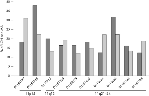
Results: Microsatellite markers D11S1758 from 11p13-15 and D11S925 from 11q23.3-24 had the highest frequency (38% and 32%, respectively) of loss of heterozygosity among all the markers analysed. Allelic loss at the marker D11S925 was seen in both leukoplakia and in all stages of HNSCC tumour tissues suggesting that it is an early event in HNSCC tumorigenesis. Microsatellite size alteration was also found to be high (> 20%) in several markers. In leukoplakia samples microsatellite instability was seen at a higher frequency than loss of the allele, indicating such alterations might initiate the process of tumorigenesis in HNSCC.

Conclusions: The high rate of chromosomal alterations at 11q21-24 in HNSCC suggests the presence of a putative tumour suppressor gene in this region.


Figures

Figure 1
Figure 1
Allelic imbalance data of the chromosome 11 markers used according to tumour type. Patients are arranged according to stage and site of the tumour. Markers with their cytogenetic positions are shown in columns at the left hand side of the figure. LMA, loss of one allele and size alteration of the other; LOH, loss of heterozygosity; MA, microsatellite size alteration; NI, non-informative; RH, retention of heterozygosity; U, unknown.
Figure 2
Figure 2
Representative autoradiographs showing loss of heterozygosity and microsatellite size alteration at different markers on chromosome 11. The markers (shown at the bottom of each panel) were amplified from paired tumour (T) and normal (N) tissues taken from selected patients (shown at the top of each panel), analysed by denaturing 6% polyacrylamide gel electrophoresis, and autoradiographed. (A) The samples from patient 538 show the retention of both alleles; the samples from patients 1367 and 3037 show loss of the lower and upper alleles, respectively. (B) The samples from patient 5090 show loss of the lower allele and a shift in the upper allele in the tumour tissue. The samples from patients 1552 and 1864 show a shift in the upper and lower allele, respectively. The arrows indicate the loss of an allele and the arrowheads indicate a shift of the allele.
Figure 3
Figure 3
Frequency of loss of heterozygosity (LOH) and microsatellite size alteration (MA) at different markers on chromosome 11 in primary head and neck squamous cell carcinomas. The dark bars indicate LOH and the lighter bars indicate MA. The names of the markers and their cytogenetic positions are shown at the bottom of each histogram.
Figure 4
Figure 4
The frequency of type of allelic imbalance in head and neck squamous cell carcinomas. Bars represent the percentage of head and neck squamous cell carcinoma tumours (HNSCC) exhibiting only loss of heterozygosity (LOH), only microsatellite size alteration (MA), or both LOH and MA in at least one marker. Each tumour was genotyped with eight to 10 markers.
Figure 5
Figure 5
Frequency of loss of heterozygosity (LOH) and microsatellite size alteration (MA) at different markers on chromosome 11 in leukoplakia. The darker bars show the percentage of LOH and the lighter bars show the percentage of MA.
Figure 6
Figure 6
Allelic imbalance data of the chromosome 11 markers in leukoplakia samples. Leukoplakia samples are arranged according to the stage of dysplasia. Markers with their cytogenetic positions are shown in columns at the left hand side of the figure. LOH, loss of heterozygosity; MA, microsatellite size alteration; NI, non-informative; RH, retention of heterozygosity.

Similar articles

Cited by

References

    1. Parkin DM, Pisani P, Ferlay J, et al. Estimates of the worldwide incidence of eighteen major cancers in 1985. Int J Cancer 1993;54:594–600. - PubMed
    1. Saranath D, Bhoite LT, Deo MG, et al. Molecular lesion in human oral cancer: the Indian scene, oral oncology. Eur J Cancer 1993;29:107–12. - PubMed
    1. Wu CL, Solan P, Thakker N, et al. Deletion mapping on the short arm of chromosome 3 in squamous cell carcinoma of the oral cavity. Cancer Res 1994;54:6484–8. - PubMed
    1. Sauders WS, Shuster S, Huang X, et al. Chromosomal instability and cytoskeletal defects in oral cancer cells. Proc Natl Acad Sci U S A 2000;97:303–8. - PMC - PubMed
    1. Van Dyke D, Worsham MJ, Beninger MS, et al. Recurrent cytogenetic abnormalities in squamous cell carcinomas of the head and neck region. Genes Chromosomes Cancer 1994;9:192–206. - PubMed

Publication types