

Review
doi: 10.1136/jcp.56.7.548.
A rare case of intracranial metastatic amelanotic melanoma with cyst
Affiliations
- PMID: 12835304
- PMCID: PMC1770002
- DOI: 10.1136/jcp.56.7.548
Item in Clipboard
Review
A rare case of intracranial metastatic amelanotic melanoma with cyst
J Clin Pathol.
2003 Jul.
Abstract
A rare case of intracranial metastatic amelanotic melanoma with cyst is presented. The patient was a 51 year old woman with a malignant melanoma arising on her right chest. Two years after a wide excision, skin and brain metastasis occurred. Brain magnetic resonance images demonstrated a tumour with a cyst in the left occipital lobe. Because the tumour showed low intensity on T1 weighted images and high intensity on T2 weighted images, the metastatic melanoma was identified as an amelanotic melanoma. Intracranial amelanotic melanoma is very rare, and there have been few reports of melanoma with cyst.
Figures
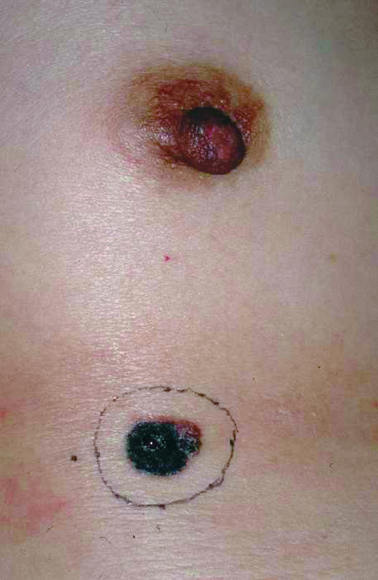
Preoperative view: the nodule was flat, irregular in shape, and 1.5 × 1.3 × 2 cm in size. An excisional biopsy with a 5 mm normal skin margin was performed to decide on the specific treatment plan.
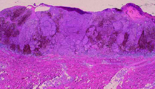
Pathological findings: the tumour cells had clearly invaded between the collagen bundles of the reticular dermis with nest formation (haematoxylin and eosin stain; original magnification, ×2).
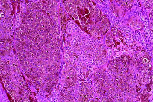
Pathological findings (haematoxylin and eosin stain; original magnification, ×20).
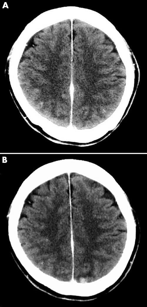
(A) A brain computed tomography (CT) image at one year after a wide excision. There are no specific findings. (B) A brain CT image at two years after a wide excision. The image shows a high density area in the left occipital lobe.
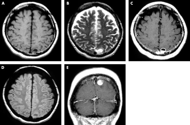
Brain magnetic resonance images at two years after a wide excision. (A) T1 weighted image (T1WI): axial view; (B) T2 weighted image (T2WI): axial view; (C) enhanced T1WI: axial view; (D) fluid attenuated inversion recovery: axial view; (E) enhanced T1WI: coronal view.
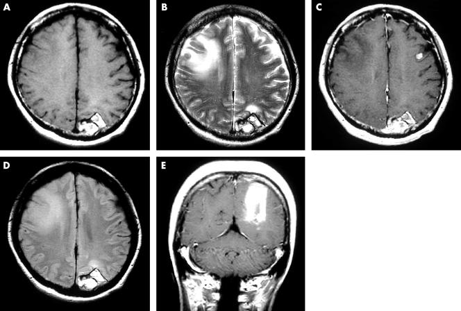
Brain magnetic resonance images at two years and one month after a wide excision. (A) T1 weighted image (T1WI): axial view; (B) T2 weighted image (T2WI): axial view; (C) enhanced T1WI: axial view; (D) fluid attenuated inversion recovery: axial view; (E) enhanced T1WI: coronal view.
Similar articles
-
A cystic amelanotic melanoma metastasis to the brain: case report.Neurocirugia (Astur). 2008 Aug;19(4):365-7. doi: 10.1016/s1130-1473(08)70225-2. Neurocirugia (Astur). 2008. PMID: 18726049
-
[A case of cystic metastatic intracranial amelanotic melanoma--analysis of findings in CT and MRI].No To Shinkei. 1990 Nov;42(11):1031-4. No To Shinkei. 1990. PMID: 2076346 Japanese.
-
Intracranial metastatic melanoma: correlation between MR imaging characteristics and melanin content.AJR Am J Roentgenol. 1995 Dec;165(6):1503-12. doi: 10.2214/ajr.165.6.7484597. AJR Am J Roentgenol. 1995. PMID: 7484597
-
[Mandibular metastasis of a cutaneous melanoma or metachronous amelanotic melanoma of the oral cavity? A case report and literature review].Ann Chir Plast Esthet. 2014 Aug;59(4):276-9. doi: 10.1016/j.anplas.2014.01.002. Epub 2014 Feb 3. Ann Chir Plast Esthet. 2014. PMID: 24503521 Review. French.
-
Amelanotic Meningeal Melanoma with Leptomeningeal Dissemination: A Case Report and Systematic Literature Review.World Neurosurg. 2019 Feb;122:229-239. doi: 10.1016/j.wneu.2018.10.199. Epub 2018 Nov 4. World Neurosurg. 2019. PMID: 30404049
Cited by
-
Intracranial amelanotic melanoma: a case report with literature review.World J Surg Oncol. 2015 May 12;13:182. doi: 10.1186/s12957-015-0600-z. World J Surg Oncol. 2015. PMID: 25963017 Free PMC article. Review.
-
A Unique Case of Intracranial Amelanotic Melanoma with BRAF V600E Mutation Successfully Treated via Molecular-targeted Therapy.NMC Case Rep J. 2023 Mar 24;10:67-73. doi: 10.2176/jns-nmc.2022-0227. eCollection 2023. NMC Case Rep J. 2023. PMID: 37065875 Free PMC article.
-
Recognizing Recurrence: History Over Symptoms in Metastatic Melanoma.Cureus. 2025 Mar 13;17(3):e80532. doi: 10.7759/cureus.80532. eCollection 2025 Mar. Cureus. 2025. PMID: 40225542 Free PMC article.
-
Intra-orbital malignant melanoma: role of MR imaging (a case report and literature review).Glob J Health Sci. 2011 Dec 29;4(1):253-8. doi: 10.5539/gjhs.v4n1p253. Glob J Health Sci. 2011. PMID: 22980115 Free PMC article. Review.
-
Amelanotic Melanoma in the Vicinity of Acquired Melanocytic Nevi and not Arising from Agminated Melanocytic Nevi: Masquerading as Pyogenic Granuloma.Indian J Dermatol. 2016 Jan-Feb;61(1):122. doi: 10.4103/0019-5154.174135. Indian J Dermatol. 2016. PMID: 26955141 Free PMC article.
References
-
- Patel JK, Didolkar MS, Pickren JW, et al. Metastatic pattern of malignant melanoma. A study of 216 autopsy cases. Am J Surg 1978;135:807–10. - PubMed
-
- Isiklar I, Leeds NE, Fuller GN, et al. Intracranial metastatic melanoma: correlation between MR imaging characteristics and melanin content. Am J Roentgenol 1995;165:1503–12. - PubMed
-
- Takahashi I, Sugimoto S, Nunomura M, et al. A case of cystic metastatic intracranial amelanotic melanoma—analysis of findings in CT and MRI. No to Shinkei 1990;42:1031–4. - PubMed
-
- Hahimoto A, Aoki M, Kawana S. Two cases of amelanotic malignant melanoma. Nishinihon Journal of Dermatology 2002;64:45–7.
Publication types
MeSH terms
LinkOut - more resources
Full Text Sources
Medical