. 2003 Jul 7;162(1):37-46.
doi: 10.1083/jcb.200303047. Epub 2003 Jun 30.

Changes in histone acetylation during mouse oocyte meiosis

Affiliations

Changes in histone acetylation during mouse oocyte meiosis

Jin-Moon Kim et al. J Cell Biol. .

Abstract

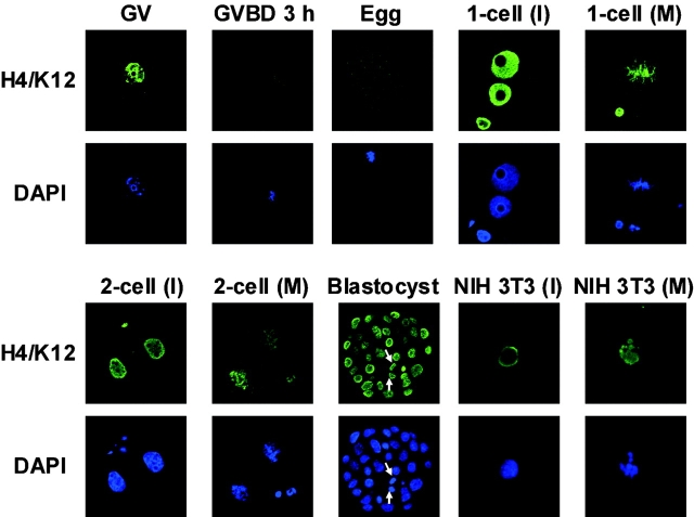
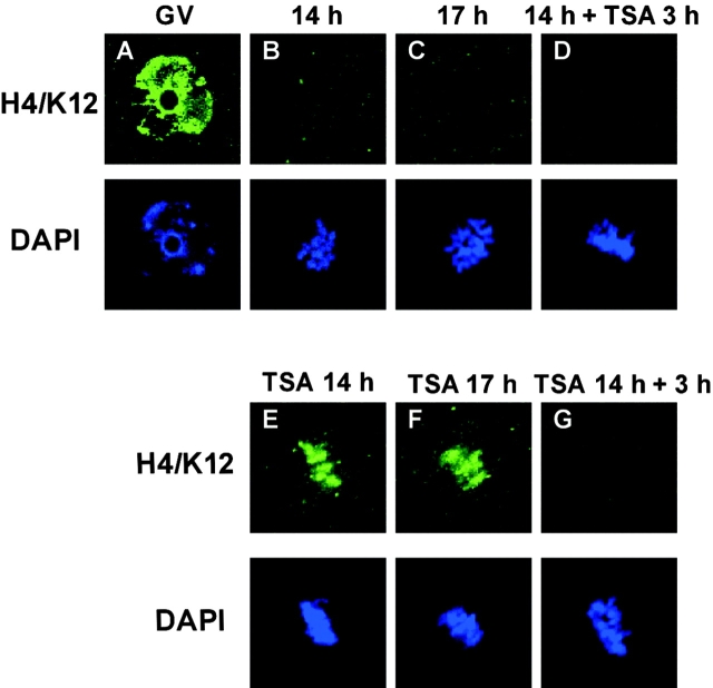
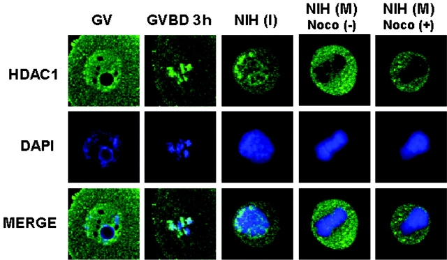
We examined global changes in the acetylation of histones in mouse oocytes during meiosis. Immunocytochemistry with specific antibodies against various acetylated lysine residues on histones H3 and H4 showed that acetylation of all the lysines decreased to undetectable or negligible levels in the oocytes during meiosis, whereas most of these lysines were acetylated during mitosis in preimplantation embryos and somatic cells. When the somatic cell nuclei were transferred into enucleated oocytes, the acetylation of lysines decreased markedly. This type of deacetylation was inhibited by trichostatin A, a specific inhibitor of histone deacetylase (HDAC), thereby indicating that HDAC is able to deacetylate histones during meiosis but not during mitosis. Meiosis-specific deacetylation may be a consequence of the accessibility of HDAC1 to the chromosome, because HDAC1 colocalized with the chromosome during meiosis but not during mitosis. As histone acetylation is thought to play a role in propagating the gene expression pattern to the descendent generation during mitosis, and the gene expression pattern of differentiated oocytes is reprogrammed during meiosis to allow the initiation of a new program by totipotent zygotes of the next generation, our results suggest that the oocyte cytoplasm initializes a program of gene expression by deacetylating histones.


Figures

Figure 1.
Figure 1.
Acetylation of lysine 14 on histone H3 at interphase (I) and metaphase (M). Oocytes, preimplantation embryos, and NIH 3T3 cells were immunostained with the anti–acetyl histone H3/lysine 14 (H3/K14) antibody. GV, oocytes at the GV stage; GVBD 3 h, oocytes after a 3-h incubation without IBMX in the first meiosis; Egg, oocytes at MII; 1-cell (I), one-cell embryos at interphase; 1-cell (M), one-cell embryos at the M phase; 2-cell (I), two-cell embryos at interphase; 2-cell (M), two-cell embryos at the M phase; Blastocyst, blastocyst-stage embryos; NIH 3T3 (I), NIH 3T3 cells at interphase; NIH 3T3 (M), NIH 3T3 cells at the M phase. Arrows indicate the condensed mitotic chromosomes in the blastocysts. Each sample was counterstained with DAPI to visualize the DNA. The acetylation of lysine 9 on histone H3 is shown in Fig. S4, available at http://www.jcb.org/cgi/content/full/jcb.200303047/DC1.
Figure 2.
Figure 2.
Acetylation of lysine 12 on histone H4 at interphase (I) and metaphase (M). Oocytes, preimplantation embryos, and NIH 3T3 cells were immunostained with the anti–acetyl histone H4/lysine 12 (H4/K12) antibody. GV, oocytes at the GV stage; GVBD 3 h, oocytes after a 3-h incubation without IBMX in the first meiosis; Egg, oocytes at MII; 1-cell (I), one-cell embryos at interphase; 1-cell (M), one-cell embryos at the M phase; 2-cell (I), two-cell embryos at interphase; 2-cell (M), two-cell embryos at the M phase; Blastocyst, blastocyst-stage embryos; NIH 3T3 (I), NIH 3T3 cells at interphase; NIH 3T3 (M), NIH 3T3 cells at the M phase. The arrows indicate condensed mitotic chromosomes in the blastocysts. Each sample was counterstained with DAPI to visualize the DNA. The acetylation of lysines 5, 8, and 16 on histone H4 is shown in Figs. S1–S3, respectively, available at http://www.jcb.org/cgi/content/full/jcb.200303047/DC1.
Figure 3.
Figure 3.
In situ analysis of the histone acetyltransferase and deacetylase in MII-stage oocytes. Oocytes at the GV and MII stages were immunostained with the anti–acetyl histone H4/lysine 12 (H4/K12) antibody. The GV-stage oocytes (A) were cultured in vitro for 14 h to mature into the MII stage (B) and were subsequently cultured for 3 h with TSA (D) or without TSA (C). The oocytes were matured in vitro for 14 h in TSA (E), and either washed to remove the TSA followed by culturing for 3 h (G), or cultured continuously in TSA for 3 h (F). Each sample was counterstained with DAPI to visualize the DNA. The results of analysis for histone H3/lysine 14 are shown in Fig. S5, available at http://www.jcb.org/cgi/content/full/jcb.200303047/DC1.
Figure 4.
Figure 4.
In situ analysis of the histone acetyltransferase and deacetylase in oocytes during the first meiosis. Oocytes in the first meiosis were immunostained with the anti–acetyl histone H4/lysine 12 (H4/K12) antibody. The GV-stage oocytes (A) were cultured in vitro for 4 h to induce the first meiosis (B) and subsequently cultured for 3 h with TSA (D) or without TSA (C). Oocytes that were undergoing the first meiosis after in vitro culture for 4 h in TSA (E) were either washed free of TSA and cultured for a further 3 h (G), or cultured continuously in TSA for 3 h (F). Each sample was counterstained with DAPI. The results of analysis for histone H3/lysine 14 are shown in Fig. S6, available at http://www.jcb.org/cgi/content/full/jcb.200303047/DC1.
Figure 5.
Figure 5.
Deacetylation of histones in transferred nuclei and chromosomes. The enucleated oocytes were transplanted with interphase nuclei (A) or metaphase chromosomes (B) of NIH 3T3 cells. NIH 3T3 cells were embedded in the perivitelline space of enucleated oocytes (embedded). Both during and after the electrofusion procedures, the enucleated oocytes and reconstructed embryos were treated with either 75 nM TSA (NT+TSA) or without TSA (NT). 2 h after electrofusion, the oocytes were subjected to immunostaining with the anti–acetyl histone H3/lysine 14 (H3/K14) or the anti–acetyl histone H4/lysine 12 (H4/K12) antibodies. Each sample was counterstained with DAPI. The deacetylation of lysine 9 on histone H3 and lysines 8 and 16 on histone H4 in the transplanted nucleus is shown in Fig. S7, available at http://www.jcb.org/cgi/content/full/jcb.200303047/DC1. The fluorescence signal of the antibody was quantified, and the results are shown in Fig. S8, available at http://www.jcb.org/cgi/content/full/jcb.200303047/DC1.
Figure 6.
Figure 6.
Localization of HDAC1 during meiosis and mitosis. The GV-stage oocytes (GV), oocytes that were incubated for 3 h in IBMX-free medium (GVBD 3 h), and NIH 3T3 cells at interphase (NIH [I]), natural mitotic phase (NIH [M] noco [−]), or nocodazole-arrested mitotic phase (NIH [M] noco [+]) were immunostained with the antibody against HDAC1 (top) and costained with DAPI (middle). The bottom row shows the merged images from the top and middle rows.

References

    1. Allfrey, V.G., R. Faulkner, and A.E. Mirsky. 1964. Acetylation and methylation of histones and their possible role in the regulation of RNA synthesis. Proc. Natl. Acad. Sci. USA. 51:786–794. - PMC - PubMed
    1. Aoki, F., and R.M. Schultz. 1999. DNA replication in the 1-cell mouse embryo: stimulatory effect of histone acetylation. Zygote. 7:165–172. - PubMed
    1. Aoki, F., D.M. Worrad, and R.M. Schultz. 1997. Regulation of transcriptional activity during the first and second cell cycles in the preimplantation mouse embryo. Dev. Biol. 181:296–307. - PubMed
    1. Agalioti, T., G. Chen, and D. Thanos. 2002. Deciphering the transcriptional histone acetylation code for a human gene. Cell. 111:381–392. - PubMed
    1. Arney, K.L., S. Bao, A.J. Bannister, T. Kouzarides, and A. Surani. 2002. Histone methylation defines epigenetic asymmetry in the mouse zygote. Int. J. Dev. Biol. 46:317–320. - PubMed

Publication types

MeSH terms