. 2003 Jul 7;198(1):91-8.
doi: 10.1084/jem.20030615. Epub 2003 Jun 30.

Bruton's tyrosine kinase cooperates with the B cell linker protein SLP-65 as a tumor suppressor in Pre-B cells

Affiliations

Bruton's tyrosine kinase cooperates with the B cell linker protein SLP-65 as a tumor suppressor in Pre-B cells

Rogier Kersseboom et al. J Exp Med. .

Abstract

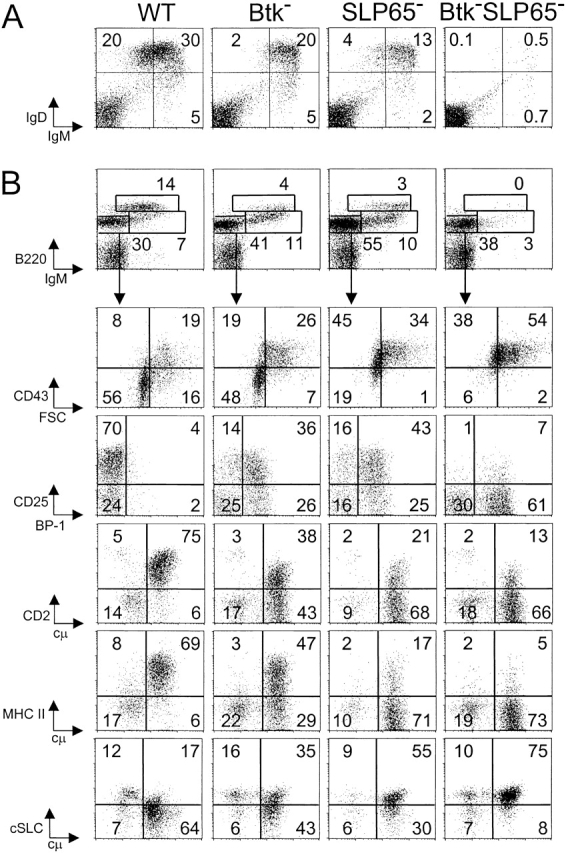
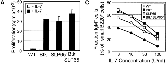
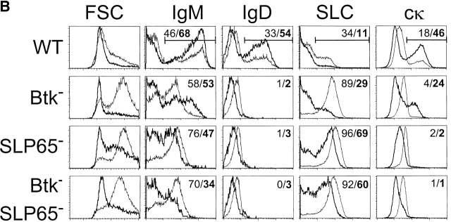
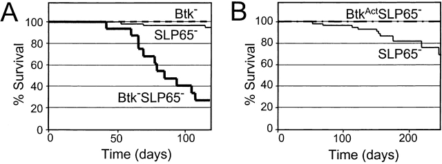
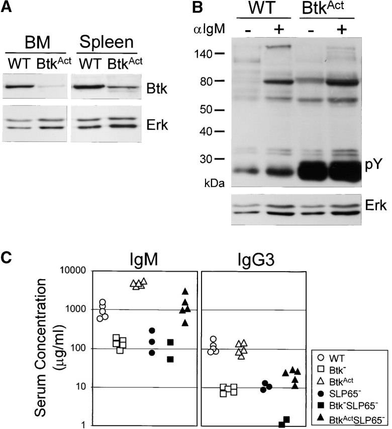
Expression of the pre-B cell receptor (pre-BCR) leads to activation of the adaptor molecule SLP-65 and the cytoplasmic kinase Btk. Mice deficient for one of these signaling proteins have an incomplete block in B cell development at the stage of large cycling pre-BCR+CD43+ pre-B cells. Our recent findings of defective SLP-65 expression in approximately 50% of childhood pre-B acute lymphoblastic leukemias and spontaneous pre-B cell lymphoma development in SLP-65-/- mice demonstrate that SLP-65 acts as a tumor suppressor. To investigate cooperation between Btk and SLP-65, we characterized the pre-B cell compartment in single and double mutant mice, and found that the two proteins have a synergistic role in the developmental progression of large cycling into small resting pre-B cells. We show that Btk/SLP-65 double mutant mice have a dramatically increased pre-B cell tumor incidence ( approximately 75% at 16 wk of age), as compared with SLP-65 single deficient mice (<10%). These findings demonstrate that Btk cooperates with SLP-65 as a tumor suppressor in pre-B cells. Furthermore, transgenic low-level expression of a constitutive active form of Btk, the E41K-Y223F mutant, prevented tumor formation in Btk/SLP-65 double mutant mice, indicating that constitutive active Btk can substitute for SLP-65 as a tumor suppressor.


Figures

Figure 1.
Figure 1.
Impaired pre-B cell maturation in Btk-deficient, SLP-65-deficient, and Btk/SLP-65 double mutant mice. (A) Flow cytometric analysis of surface IgM/IgD expression on total lymphoid cells in the spleen. (B) Expression profiles of B220 and IgM on total lymphoid cells in the BM (top). The B220+IgM pro-/pre-B cell fraction was gated and analyzed for the indicated markers (bottom). Data are displayed as dot plots and the percentages of cells within the indicated quadrants or gates are given. Data shown are representative of four mice examined within each group.
Figure 2.
Figure 2.
Analysis of IL-7 driven BM cultures from Btk and SLP-65 mutant mice. (A) Proliferative response to 100 U/ml IL-7, as determined by [3H]thymidine incorporation after 5 d of culture. Bars represent mean cpm and SEM of triplicate cultures. (B) Forward scatter (FSC) values and expression profiles of IgM, IgD, SLC, and cytoplasmic κ L chain of IL-7 driven BM cultures from the indicated mice. Data are displayed as histogram overlays of B220+ cells, either cultured under proliferating conditions (with 100 U/ml IL-7 for 7 d, thin lines) or under differentiating conditions (after 5 d of culture with IL-7 and subsequently without IL-7 for 2 d, bold lines). The percentages shown represent the fractions of the cells that are within the indicated marker under the two different culture conditions. (C) Percentage of surface IgM+ cells within the fraction of small FSC B220+ cells after 7 d of culture in the presence of the indicated concentrations of IL-7. Data are representative of four mice per group.
Figure 2.
Figure 2.
Analysis of IL-7 driven BM cultures from Btk and SLP-65 mutant mice. (A) Proliferative response to 100 U/ml IL-7, as determined by [3H]thymidine incorporation after 5 d of culture. Bars represent mean cpm and SEM of triplicate cultures. (B) Forward scatter (FSC) values and expression profiles of IgM, IgD, SLC, and cytoplasmic κ L chain of IL-7 driven BM cultures from the indicated mice. Data are displayed as histogram overlays of B220+ cells, either cultured under proliferating conditions (with 100 U/ml IL-7 for 7 d, thin lines) or under differentiating conditions (after 5 d of culture with IL-7 and subsequently without IL-7 for 2 d, bold lines). The percentages shown represent the fractions of the cells that are within the indicated marker under the two different culture conditions. (C) Percentage of surface IgM+ cells within the fraction of small FSC B220+ cells after 7 d of culture in the presence of the indicated concentrations of IL-7. Data are representative of four mice per group.
Figure 3.
Figure 3.
(A) Kaplan-Meier tumor-free survival estimates for Btk-deficient (dotted line), SLP-65−/− (thin line), and Btk-SLP-65−/− mice (bold line). Tumor-free survival in Btk-SLP-65−/− mice was significantly reduced (P < 0.0001 by log-rank) compared with SLP-65−/− mice. (B) Kaplan-Meier tumor-free survival estimates for SLP-65−/− (thin line) and BtkActSLP-65−/− mice (dotted line). Tumor-free survival in BtkActSLP-65−/− mice was significantly enhanced (P = 0.04 by log-rank) compared with SLP-65−/− mice.
Figure 4.
Figure 4.
Low-level BtkAct expression can partially substitute for the absence of Btk and SLP-65. (A) Western blotting analysis of Btk expression in WT and BtkAct B cells from BM and spleen. Membrane was reblotted with anti-Erk. (B) Protein tyrosine phosphorylation in extracts of untreated and anti-IgM stimulated WT or BtkAct splenic B cells, analyzed by immunoblotting with a phosphotyrosine (pY)-specific antibody. Membrane was reblotted with anti-Erk. (C) Serum concentrations of IgM and IgG3 in the indicated mutant mouse strains. Mice were 2 mo of ages and each symbol represents an individual animal. (D) Flow cytometric analysis of surface IgM/IgD expression on total lymphoid cells in the spleen of the indicated mice. (E) Expression profiles of B220 and IgM on total lymphoid cells in the BM of the indicated mice (top). The B220+IgM- pro-/pre-B cell fractions were gated and analyzed for CD43/FSC and cytoplasmic SLC and μ H chain (bottom). Data are displayed as dot plots and the percentages of cells within the indicated quadrants or gates are given. Data shown are representative of four mice examined within each group.
Figure 4.
Figure 4.
Low-level BtkAct expression can partially substitute for the absence of Btk and SLP-65. (A) Western blotting analysis of Btk expression in WT and BtkAct B cells from BM and spleen. Membrane was reblotted with anti-Erk. (B) Protein tyrosine phosphorylation in extracts of untreated and anti-IgM stimulated WT or BtkAct splenic B cells, analyzed by immunoblotting with a phosphotyrosine (pY)-specific antibody. Membrane was reblotted with anti-Erk. (C) Serum concentrations of IgM and IgG3 in the indicated mutant mouse strains. Mice were 2 mo of ages and each symbol represents an individual animal. (D) Flow cytometric analysis of surface IgM/IgD expression on total lymphoid cells in the spleen of the indicated mice. (E) Expression profiles of B220 and IgM on total lymphoid cells in the BM of the indicated mice (top). The B220+IgM- pro-/pre-B cell fractions were gated and analyzed for CD43/FSC and cytoplasmic SLC and μ H chain (bottom). Data are displayed as dot plots and the percentages of cells within the indicated quadrants or gates are given. Data shown are representative of four mice examined within each group.
Figure 5.
Figure 5.
Characterization of pre-B cell tumors by flow cytometry. (A) Dot plots for cytoplasmic SLC and μ H chain in gated B220+ cells from tumor samples from the indicated mice, grown for 1 to 3 wk in the presence of IL-7. (B) Flow cytometric analysis of lacZ expression in gated CD19+B220+ pre-B lymphoma cells in a lymph node from a Btk+/−SLP-65−/− mouse. (C) Phenotype of two pre-B cell lymphoma cultures, showing variable expression of cytoplasmic κ L chain, CD43, and CD2. Cell suspensions were stained for the indicated markers in combination with B220, and the results are displayed as dot plots of gated B220+ cells. (D) Phenotype of two separate tumor cell suspensions derived from BM and mesenteric lymph node from a single mouse, which were cultured in the presence of Il-7 for 7 d.

References

    1. Meffre, E., R. Casellas, and M.C. Nussenzweig. 2000. Antibody regulation of B cell development. Nat. Immunol. 1:379–385. - PubMed
    1. Melchers, F., E. ten Boekel, T. Seidl, X.C. Kong, T. Yamagami, K. Onishi, T. Shimizu, A.G. Rolink, and J. Andersson. 2000. Repertoire selection by pre-B-cell receptors and B-cell receptors, and genetic control of B-cell development from immature to mature B cells. Immunol. Rev. 175:33–46. - PubMed
    1. Conley, M.E., J. Rohrer, L. Rapalus, E.C. Boylin, and Y. Minegishi. 2000. Defects in early B-cell development: comparing the consequences of abnormalities in pre-BCR signaling in the human and the mouse. Immunol. Rev. 178:75–90. - PubMed
    1. Shaffer, A.L., and M.S. Schlissel. 1997. A truncated heavy chain protein relieves the requirement for surrogate light chains in early B cell development. J. Immunol. 159:1265–1275. - PubMed
    1. Rolink, A.G., T. Winkler, F. Melchers, and J. Andersson. 2000. Precursor B cell receptor-dependent B cell proliferation and differentiation does not require the bone marrow or fetal liver environment. J. Exp. Med. 191:23–32. - PMC - PubMed

Publication types

MeSH terms