

Review
doi: 10.1128/JB.185.14.4022-4030.2003.
Homotrimeric, beta-stranded viral adhesins and tail proteins
Affiliations
- PMID: 12837775
- PMCID: PMC164894
- DOI: 10.1128/JB.185.14.4022-4030.2003
Item in Clipboard
Review
Homotrimeric, beta-stranded viral adhesins and tail proteins
J Bacteriol.
2003 Jul.
No abstract available
Figures
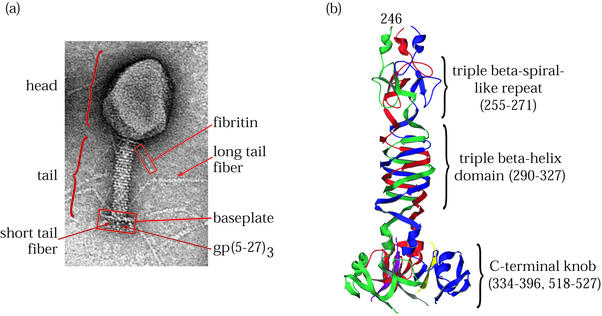
T4 virion and its short tail fiber, gp12. (a) Electron micrograph of bacteriophage T4 showing the locations of structural proteins and features. (b) Ribbon diagram of the T4 short tail fiber structure (67). The C-terminal domain at the bottom of the figure binds irreversibly to the bacterial host cell LPS. Residues 290 to 327 comprise a triple β-helix. A single β-strand motif similar but not identical to a triple β-spiral repeat is seen near the N terminus of the structure at the top. The three subunits are shown in red, green, and blue. (C-terminal strands whose connectivities were not assigned are shown in yellow, purple, and gray.)
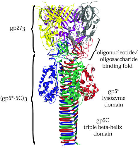
T4 tail lysozyme complex (gp27-gp5). Shown is a ribbon diagram of the lysozyme complex structure (33), which contains three copies each of gp27 and gp5. In the mature protein, gp5 is proteolytically cleaved, yielding gp5* and gp5C, which both remain associated with the complex. The three gp5 subunits, composed of the gp5* and gp5C fragments, are shown in red, green, and blue, and the three gp27 subunits are shown in yellow, purple, and gray. Residues of positions 436 to 575 of gp5 are found in the gp5C fragment and form a remarkable seven-turn, triple β-helix.
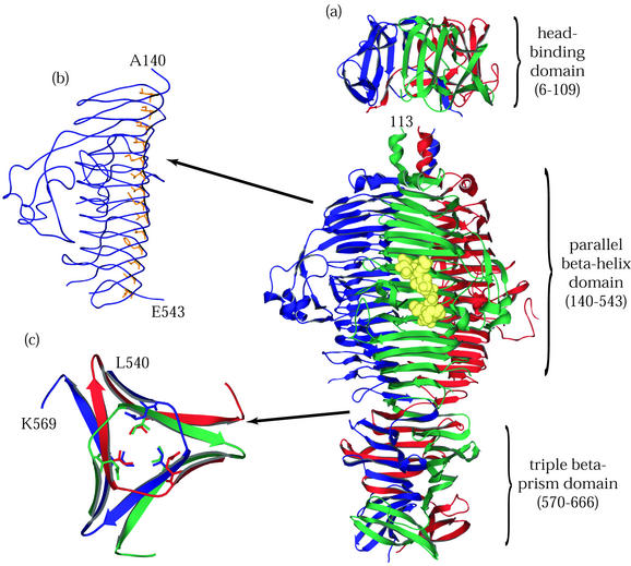
P22 tailspike protein. The illustrations are based on structures determined by Steinbacher and coworkers (59-62). (a) The entire P22 tailspike protein, shown bound to the nonasaccharide from S. enterica serovar 253Ty O-antigen (in yellow space-filling representation). The N-terminal domain is at the top, and the three subunit chains are shown in red, green, and blue. (b) An interior hydrophobic stack from one of the three identical single-chain, parallel β-helices is shown with side chains highlighted in yellow. (c) Residues 540 to 569, viewed from above and showing inwardly pointing hydrophobic residues. This region, which spans the interdigitated domain, forms one turn of a triple-stranded β-helix and is involved in trimer stability.
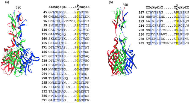
Viral attachment fibers that have triple β-spiral repeats. (a) Structure of the human Ad2 penton fiber (65) showing four triple β-spiral repeats (residues 320 to 392) in the N-terminal shaft domain and the CAR binding C-terminal knob (residues 399 to 582). (b) Model of the reovirus σ1 attachment protein structure (15) showing three triple β-spiral repeats (residues 246 to 309) and the C-terminal knob (residues 310 to 455). The two knob proximal repeats are separated by a flexible spacer. The knob is a β-barrel composed of two key motifs shown as Greek letters. Both complexes are shown with the N-terminal domains at the top and the chains colored red, green, and blue. For both proteins, alignments of previously defined 15-amino-acid repeats are shown, conserved hydrophobic or structural residues are highlighted, and a canonical repeat motif is given at the top of each alignment.
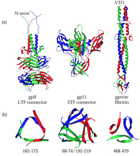
T4 tail proteins. (a) Ribbon diagrams of gp9, gp11, and gpwac from structures determined by Kostyuchenko et al. (38), Tao et al. (64), and Leiman et al. (41), respectively. gp9 connects the long tail fibers to the baseplate. gp11 connects the short tail fibers to the baseplate. Fibritin, the wac gene product, is found in the virus as a long “whisker” attached to the T4 neck and plays a role in long tail fiber assembly and retraction. Ribbon diagrams are displayed with N-terminal domains at the top and monomers in red, green, and blue. (b) Ribbon diagrams of the β-annulus domains displayed below the diagrams of the proteins in which they are found. Each annular domain is a cross section of the protein viewed from above.
References
-
- Baker, K. A., R. E. Dutch, R. A. Lamb, and T. S. Jardetzky. 1999. Structural basis for paramyxovirus-mediated membrane fusion. Mol. Cell 3:309-319. - PubMed
-
- Barton, E. S., J. C. Forrest, J. L. Connolly, J. D. Chappell, Y. Liu, F. J. Schnell, A. Nusrat, C. A. Parkos, and T. S. Dermody. 2001. Junction adhesion molecule is a receptor for reovirus. Cell 104:441-451. - PubMed
-
- Bergelson, J. M., J. A. Cunningham, G. Droguett, E. A. Kurt-Jones, A. Krithivas, J. S. Hong, M. S. Horwitz, R. L. Crowell, and R. W. Finberg. 1997. Isolation of a common receptor for Coxsackie B viruses and adenoviruses 2 and 5. Science 275:1320-1323. - PubMed
-
- Betts, S., and J. King. 1999. There's a right way and a wrong way: in vivo and in vitro folding, misfolding and subunit assembly of the P22 tailspike. Struct. Fold Des. 7:R131-R139. - PubMed
Publication types
MeSH terms
Substances
Grants and funding
LinkOut - more resources
Full Text Sources
Other Literature Sources
