. 2003 Jul;185(14):4031-7.
doi: 10.1128/JB.185.14.4031-4037.2003.

Crystal structure of the YchF protein reveals binding sites for GTP and nucleic acid

Affiliations

Crystal structure of the YchF protein reveals binding sites for GTP and nucleic acid

Alexey Teplyakov et al. J Bacteriol. 2003 Jul.

Abstract

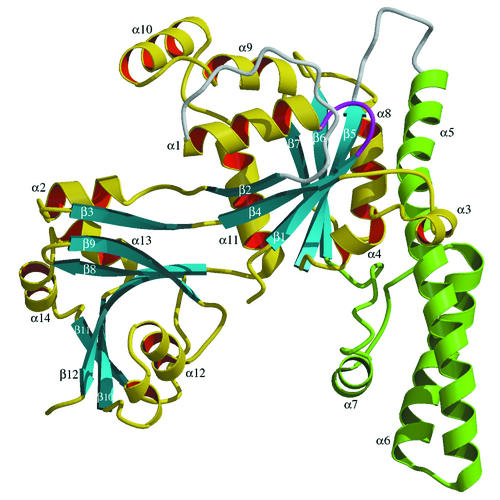
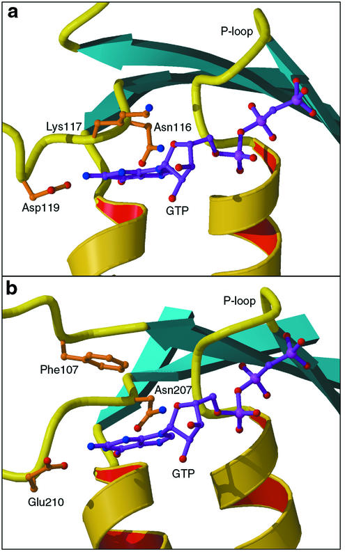
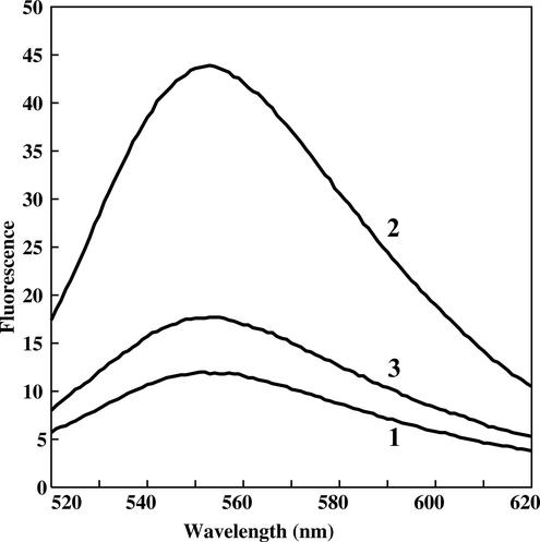
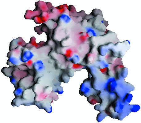
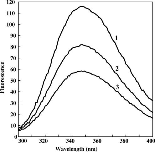
The bacterial protein encoded by the gene ychF is 1 of 11 universally conserved GTPases and the only one whose function is unknown. The crystal structure determination of YchF was sought to help with the functional assignment of the protein. The YchF protein from Haemophilus influenzae was cloned and expressed, and the crystal structure was determined at 2.4 A resolution. The polypeptide chain is folded into three domains. The N-terminal domain has a mononucleotide binding fold typical for the P-loop NTPases. An 80-residue domain next to it has a pronounced alpha-helical coiled coil. The C-terminal domain features a six-stranded half-barrel that curves around an alpha-helix. The crablike three-domain structure of YchF suggests the binding site for a double-stranded nucleic acid in the cleft between the domains. The structure of the putative GTP-binding site is consistent with the postulated guanine specificity of the protein. Fluorescence measurements have demonstrated the ability of YchF to bind a double-stranded nucleic acid and GTP. Taken together with other experimental data and genomic analysis, these results suggest that YchF may be part of a nucleoprotein complex and may function as a GTP-dependent translation factor.


Figures

FIG. 1.
FIG. 1.
Ribbon presentation of the polypeptide fold of YchF. The α-helical domain is shown in green. Disordered loops are shown in white. The NTP-binding P-loop is shown in purple. The figure was produced with Molscript (18) and Raster3D (23).
FIG. 2.
FIG. 2.
Amino acid sequence of the H. influenzae YchF in one-letter code. Residues conserved in all family members with no more than one exception are enclosed in boxes. Residues with conservative replacements in two or three family members are in bold letters. Secondary structure elements calculated with DSSP (16) are indicated above the sequence.
FIG. 3.
FIG. 3.
Guanine recognition site in Ras (Protein Data Bank entry 121P), a typical G protein (a), and YchF (b). GTP is shown in purple, as observed in Ras and as modeled in YchF. The lysine residue of the consensus sequence NKXD (Lys117 in Ras) is spatially replaced with Phe107 in YchF.
FIG. 4.
FIG. 4.
Fluorescence of TNP-GTP in the absence and presence of GTP. The protein was present at a concentration of 2.5 μM. The increase in the fluorescence intensity of TNP-GTP with respect to that in the protein-free solution is plotted over the TNP-GTP concentration. The fluorescence intensity was recorded at 25°C at a wavelength of 555 nm, and the excitation wavelength was 408 nm.
FIG. 5.
FIG. 5.
Fluorescence spectra of TNP-GTP in the absence and presence of YchF and Mg2+. Line 1, 2.5 μM TNP-GTP; line 2, 2.5 μM TNP-GTP plus 2.5 μM YchF; line 3, 2.5 μM TNP-GTP plus 2.5 μM YchF plus 2 mM Mg2+. The emission spectrum was recorded at 25°C, and the excitation wavelength was 408 nm.
FIG. 6.
FIG. 6.
Electrostatic surface potential calculated with GRASP (28). Positive charges are blue, and negative charges are red. The orientation of the molecule is as shown in Fig. 1.
FIG. 7.
FIG. 7.
Quenching of fluorescence of tryptophan upon addition of dsDNA to the YchF protein. The protein concentration was 100 nM. Line 1, no dsDNA; line 2, 50 nM (per base pair) dsDNA; line 3, 100 nM (per base pair) dsDNA.

References

    1. Bateman, A., E. Birney, R. Durbin, S. R. Eddy, K. L. Howe, and E. L. Sonnhammer. 2000. The Pfam protein families database. Nucleic Acids Res. 28:263-266. - PMC - PubMed
    1. Berman, H. M., J. Westbrook, Z. Feng, G. Gilliland, T. N. Bhat, H. Weissig, I. N. Shindyalov, and P. E. Bourne. 2000. The Protein Data Bank. Nucleic Acids Res. 28:235-242. - PMC - PubMed
    1. Biou, V., A. Yaremchuk, M. Tukalo, and S. Cusack. 1994. The 2.9 Å crystal structure of T. thermophilus seryl-tRNA synthetase complexed with tRNASer. Science 263:1404-1410. - PubMed
    1. Bourne, H. R., D. A. Sanders, and F. McCormick. 1990. The GTPase superfamily: a conserved switch for diverse cell functions. Nature 348:125-132. - PubMed
    1. Bourne, H. R., D. A. Sanders, and F. McCormick. 1991. The GTPase superfamily: conserved structure and molecular mechanism. Nature 349:117-127. - PubMed

Publication types

Associated data

LinkOut - more resources