. 2003 Jul;185(14):4074-80.
doi: 10.1128/JB.185.14.4074-4080.2003.

A novel adenylate binding site confers phosphopantetheine adenylyltransferase interactions with coenzyme A

Affiliations

A novel adenylate binding site confers phosphopantetheine adenylyltransferase interactions with coenzyme A

Tina Izard. J Bacteriol. 2003 Jul.

Abstract

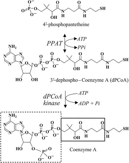
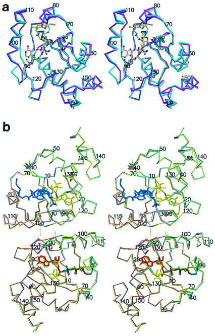
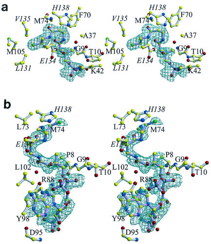
Phosphopantetheine adenylyltransferase (PPAT) regulates the key penultimate step in the essential coenzyme A (CoA) biosynthetic pathway. PPAT catalyzes the reversible transfer of an adenylyl group from Mg(2+):ATP to 4'-phosphopantetheine to form 3'-dephospho-CoA (dPCoA) and pyrophosphate. The high-resolution crystal structure of PPAT complexed with CoA has been determined. Remarkably, CoA and the product dPCoA bind to the active site in distinct ways. Although the phosphate moiety within the phosphopantetheine arm overlaps, the pantetheine arm binds to the same pocket in two distinct conformations, and the adenylyl moieties of these two ligands have distinct binding sites. Moreover, the PPAT:CoA crystal structure confirms the asymmetry of binding to the two trimers within the hexameric enzyme. Specifically, the pantetheine arm of CoA bound to one protomer within the asymmetric unit displays the dPCoA-like conformation with the adenylyl moiety disordered, whereas CoA binds the twofold-related protomer in an ordered and unique fashion.


Figures

FIG. 1.
FIG. 1.
The final two steps in the biosynthetic pathway of CoA. CoA biosynthesis occurs in a series of five steps that utilize pantothenate (vitamin B5), cysteine, and ATP. PPAT catalyzes the penultimate step, the reversible transfer of an adenylyl group from ATP to 4′-phosphopantetheine to form dPCoA and pyrophosphate. Subsequent phosphorylation at the 3′-hydroxyl of the ribose ring by dPCoA kinase produces the acyl group carrier, CoA. For CoA, the 3′-phosphate ADP is shown by a dashed box, the pantothenic acid group of pantetheine is outlined by the solid box, and the β-mercaptoethylamine of pantetheine is not boxed.
FIG. 2.
FIG. 2.
(a) Stereodiagram of the PPAT:CoA Cα trace of the two superimposed subunits within the asymmetric unit. Protomer A showing the entire inhibitor (magenta) and the twofold-related superimposed protomer B (teal) are shown. Ligands are shown in ball and stick representation. Oxygen (red) nitrogen (blue), sulfur (green), carbon (yellow), and phosphorus (black) atoms are shown. The overall rms deviation for the 314 equivalent Cα positions of the superposition of the two subunits in the asymmetric unit is 0.81 Å. This color coding of atoms is also used in panel b and in Fig. 3. (b) Stereodiagram of the PPAT Cα trace of the dimer within the asymmetric unit bound to CoA (blue), dPCoA (red) (Protein Data Bank identification code [PDB ID] 1B6T) (14), ATP (yellow) (PDB ID 1GN8) (16), and Ppant (green) (PDB ID 1QJC) (16). Protomer A (top subunit) binds CoA in the PPAT:CoA structure and ATP in the PPAT:ATP structure, while protomer B (bottom subunit) binds dPCoA (PPAT:dPCoA structure), ATP (PPAT:ATP structure), and Ppant (PPAT:Ppant structure) but shows only partially ordered binding to CoA (PPAT:CoA structure). This figure was produced with MolScript (19) or BobScript (11) and Raster3D (22) programs.
FIG. 3.
FIG. 3.
Stereo views of the binding of CoA to PPAT. For clarity, water molecules (red spheres) are not labeled, and only the groups that participate in binding (i.e., main chain or the side chain atoms) are shown. The protein structures shown in panels a and b are in similar orientations. Labels for residues belonging to a threefold-related subunit are italicized. Final σA (28) weighted difference omit electron density map for CoA bound to subunit B (a), where electron density is visible only for the Ppant moiety of CoA, and the twofold-related protomer A (b). The contour level of the electron density maps is 2.5σ, and the resolution is 1.78 Å. The bonds of CoA (pink) and the bonds of the enzyme (white) are shown. Color coding of atoms is explained in the legend to Fig. 2. This figure was produced with MolScript (19) or BobScript (11) and Raster3D (22) programs.

References

    1. Aghajanian, S., and D. M. Worrall. 2002. Identification and characterization of the gene encoding the human phosphopantetheine adenylyltransferase and dephospho-CoA kinase bifunctional enzyme (CoA synthase). Biochem. J. 365:13-18. - PMC - PubMed
    1. Bork, P., L. Holm, E. V. Koonin, and C. Sander. 1995. The cytidylyltransferase superfamily: identification of the nucleotide-binding site and fold prediction. Proteins 22:259-266. - PubMed
    1. Brünger, A. T., J. Kuriyan, and M. Karplus. 1988. Crystallographic R-factor refinement by molecular dynamics. Science 235:458-460. - PubMed
    1. Brünger, A. T., P. D. Adams, G. M. Clore, W. L. DeLano, P. Gros, R. W. Grosse-Kunstleve, J.-S. Jiang, J. Kunszeweski, N. Nilges, M. S. Pannu, R. J. Read, L. M. Rice, T. Simonson, and G. L. Warren. 1998. Crystallography & NMR system: a new software suite for macromolecular structure determination. Acta Crystallogr. Sect. D 54:905-921. - PubMed
    1. Brünger, A. T. 1992. Free R value: a novel statistical quantity for assessing the accuracy of crystal structures. Nature 355:472-475. - PubMed

Publication types

Substances

Associated data

LinkOut - more resources