. 2003 Jul;69(7):3739-50.
doi: 10.1128/AEM.69.7.3739-3750.2003.

Characterization and in situ carbon metabolism of phototrophic consortia

Affiliations

Characterization and in situ carbon metabolism of phototrophic consortia

Jens Glaeser et al. Appl Environ Microbiol. 2003 Jul.

Abstract

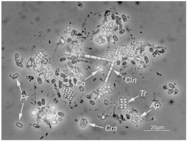
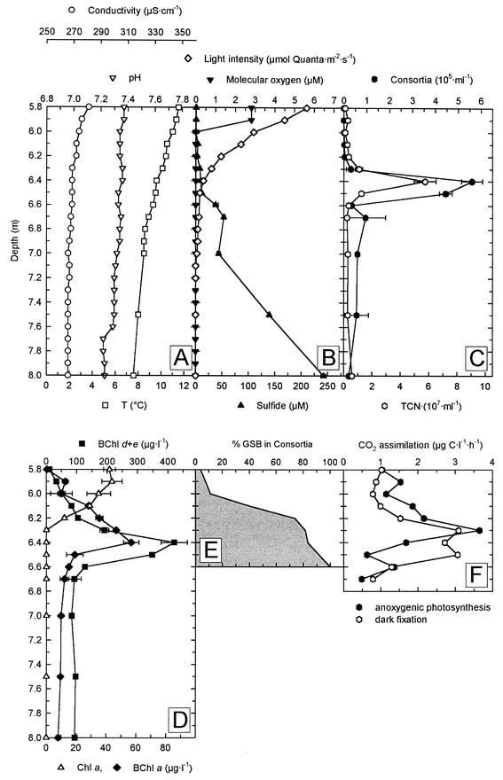
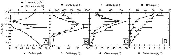
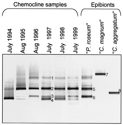
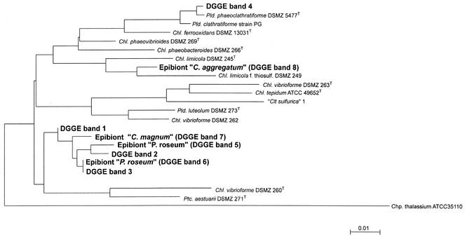
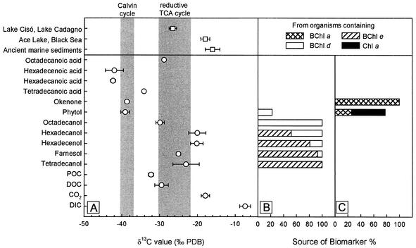
A dense population of the phototrophic consortium "Pelochromatium roseum" was investigated in the chemocline of a temperate holomictic lake (Lake Dagow, Brandenburg, Germany). Fluorescence in situ hybridization revealed that the brown epibionts of "P. roseum" constituted up to 37% of the total bacterial cell number and up to 88% of all green sulfur bacteria present in the chemocline. Specific amplification of 16S rRNA gene fragments of green sulfur bacteria and denaturing gradient gel electrophoresis fingerprinting yielded a maximum of four different DNA bands depending on the year of study, indicating that the diversity of green sulfur bacteria was low. The 465-bp 16S rRNA gene sequence of the epibiont of "P. roseum" was obtained after sorting of individual consortia by micromanipulation, followed by a highly sensitive PCR. The sequence obtained represents a new phylotype within the radiation of green sulfur bacteria. Maximum light-dependent H(14)CO(3)(-) fixation in the chemocline in the presence of 3-(3,4-dichlorophenyl)-1,1-dimethylurea suggested that there was anaerobic autotrophic growth of the green sulfur bacteria. The metabolism of the epibionts was further studied by determining stable carbon isotope ratios (delta(13)C) of their specific biomarkers. Analysis of photosynthetic pigments by high-performance liquid chromatography revealed the presence of high concentrations of bacteriochlorophyll (BChl) e and smaller amounts of BChl a and d and chlorophyll a in the chemocline. Unexpectedly, isorenieratene and beta-isorenieratene, carotenoids typical of other brown members of the green sulfur bacteria, were absent. Instead, four different esterifying alcohols of BChl e were isolated as biomarkers of green sulfur bacterial epibionts, and their delta(13)C values were determined. Farnesol, tetradecanol, hexadecanol, and hexadecenol all were significantly enriched in (13)C compared to bulk dissolved and particulate organic carbon and compared to the biomarkers of purple sulfur bacteria. The difference between the delta(13)C values of farnesol, the major esterifying alcohol of BChl e, and CO(2) was -7.1%, which provides clear evidence that the mode of growth of the green sulfur bacterial epibionts of "P. roseum" in situ is photoautotrophic.


Figures

FIG. 1.
FIG. 1.
Phase-contrast microphotograph of the bacterial community present at a depth of 6.4 m of in Lake Dagow on 4 August 1998. Ap, Amoebobacter purpureus-like cells; Cln, Chloronema-like cells; Cm, “C. magnum”; Co, Chromatium okenii-like cells; Pr, “P. roseum”; Tr, Thiopedia rosea-like cells. With bright-field microscopy, “P. roseum” has a distinct brown color.
FIG. 2.
FIG. 2.
Physical, chemical, and biological parameters in the chemocline of Lake Dagow on 4 July 1998. (A) Conductivity, temperature, and pH. (B) Concentrations of molecular oxygen and sulfide. (C) Numbers of phototrophic consortia and total cell number (TCN). (D) Concentrations of BChl a, BChl d and e, and Chl a. (E) Fraction of green sulfur bacteria (GSB) associated with phototrophic consortia as determined by fluorescence in situ hybridization. (F) CO2 assimilation rates as determined by incubation with NaH14CO3.
FIG. 3.
FIG. 3.
Composition of phototrophic bacteria in the Lake Dagow chemocline on 19 July 1999 as determined by HPLC analyses. (A) Numbers of phototrophic consortia, molecular oxygen saturation (expressed as a percentage of air saturation), and concentrations of sulfide. (B to D) Photosynthetic pigments of green sulfur bacteria (BChl d and BChl e) (B), purple sulfur bacteria (BChl a and okenone) (C), and cyanobacteria and algae (Chl a and β-carotene) (D).
FIG. 4.
FIG. 4.
16S rRNA gene fingerprints of green sulfur bacteria from the chemocline microbial community in Lake Dagow for six consecutive years compared to fingerprints of isolated consortia. The latter were generated by using 10 isolated intact consortia (central bacterium plus attached epibionts) of “C. aggregatum,” 10 consortia of “P. roseum,” and five consortia of “C. magnum.” A negative image of an ethidium bromide-stained DGGE gel is shown. The numbers of DNA bands correspond to sequences in the phylogenetic tree in Fig. 5.
FIG. 5.
FIG. 5.
Phylogenetic position of the “P. roseum” epibiont in the radiation of green sulfur bacteria. A phylogenetic tree was constructed by the maximum-likelihood method from full-length sequences of the most closely related green sulfur bacteria. The partial 16S rRNA gene sequences were then inserted by using a maximum-parsimony tool as implemented in the ARB phylogeny package. The DNA sequence of the epibiont of “P. roseum” in band 6 is identical to the DNA sequence in band 3 from the entire chemocline microbial community (compare Fig. 4). Bar = 0.01 fixed point mutation per base. Pld., Pelodictyon; Chl., Chlorobium; Clt., Clathrochloris; Ptc., Prosthecochloris; Chp., Chloroherpeton.
FIG. 6.
FIG. 6.
δ13C values and origin of biomarkers of phototrophic bacteria isolated from a depth of 6.8 m in Lake Dagow on 19 July 1999. (A) δ13C values of specific biomarkers. Symbols: ○, δ13C values from the chemocline of Lake Dagow; □, average of δ13C values from previously published papers (for Lake Cisó and Lake Cadagno, isorenieratene from sediments [20, 50]; for Ace Lake and the Black Sea, isorenieratene, chlorobactene, and farnesane from sediments [22, 54, 57]; for ancient sediments, isorenieratene isolated from rocks [19, 21, 42, 55, 56]). The shaded areas indicate the range of Δδ13C values for the biomass of green and purple sulfur bacteria for the chemocline of Lake Dagow as calculated from previously published data (29, 47, 59). TCA, tricarboxylic acid. (B) Source of specific biomarkers derived from CO2 fixation by the reversed citric acid cycle. (C) Source of specific biomarkers derived from CO2 fixation via the Calvin cycle.

References

    1. Altschul, S. F., T. L. Madden, A. A. Schäffer, J. Zhang, W. Miller, and D. J. Lipman. 1997. Gapped BLAST and PSI-BLAST: a new generation of protein database search programs. Nucleic Acids Res. 25:3389-3402. - PMC - PubMed
    1. Arellano, J. B., C. M. Borrego, A. Martínez-Planells, and L. J. Garcia-Gil. 2001. Effect of carotenoid deficiency on cells and chlorosomes of Chlorobium phaeobacteroides. Arch. Microbiol. 175:226-233. - PubMed
    1. Behrens, A., P. Schaeffer, S. Bernasconi, and P. Albrecht. 2000. Mono- and bicyclic squalene derivatives as potential proxies for anaerobic photosynthesis in lacustrine sulfur-rich sediments. Geochim. Cosmochim. Acta 64:3327-3336.
    1. Berry, J. A., J. Throughton, and O. E. Bjørkman. 1974. Carbon isotope fractionation in open and closed systems. Carnegie Inst. Wash. Year Book 73:785-790.
    1. Borrego, C. M., J. B. Arellano, C. A. Abella, T. Gillbro, and L. J. Garcia-Gil. 1999. The molar extinction coefficient of bacteriochlorophyll e and the pigment stoichiometry in Chlorobium phaeobacteroides. Photosynth. Res. 60:257-264.

LinkOut - more resources