. 2003 Jul;139(5):1041-9.
doi: 10.1038/sj.bjp.0705330.

Enhancement of lipopolysaccharide-stimulated JNK activity in rat aortic smooth muscle cells by pharmacological and adenovirus-mediated inhibition of inhibitory kappa B kinase signalling

Affiliations

Enhancement of lipopolysaccharide-stimulated JNK activity in rat aortic smooth muscle cells by pharmacological and adenovirus-mediated inhibition of inhibitory kappa B kinase signalling

Christopher J MacKenzie et al. Br J Pharmacol. 2003 Jul.

Abstract

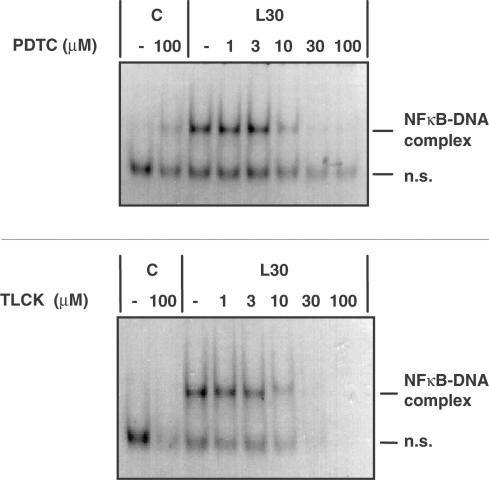
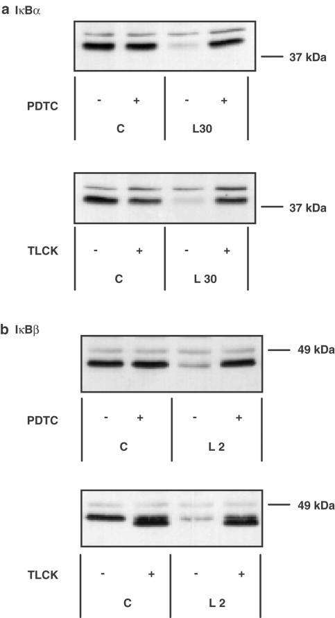
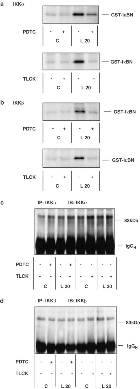
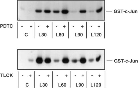
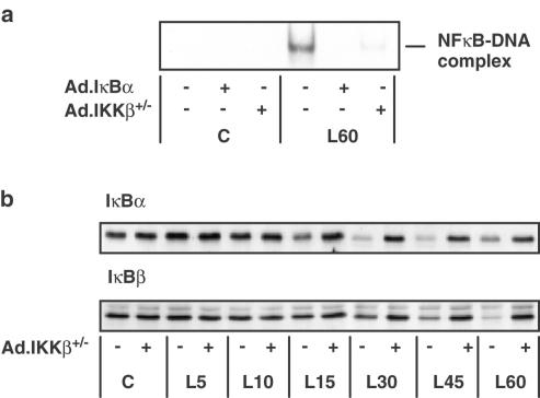
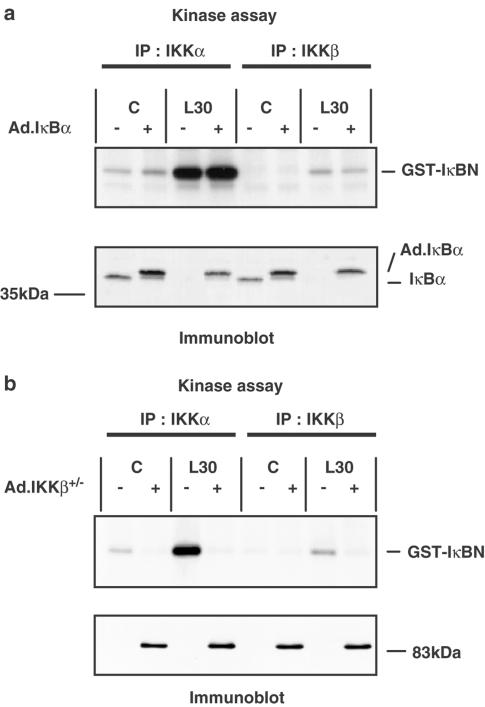
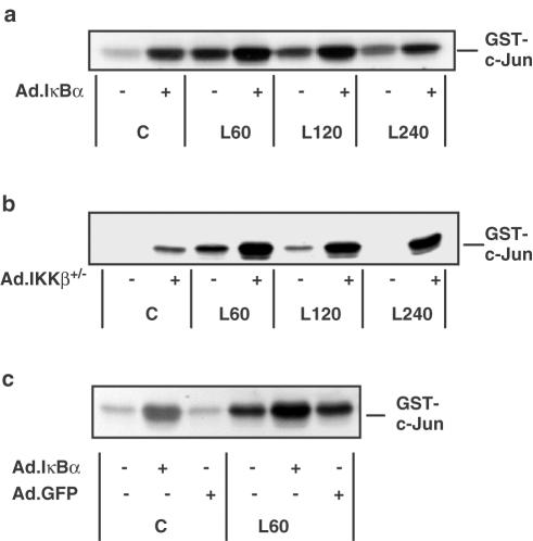
1. In rat aortic smooth muscle cells (RASMCs), the putative nuclear factor kappa B (NFkappaB) inhibitor Pyrrolidine dithiocarbamate (PDTC) was found to inhibit lipopolysaccharide (LPS)-stimulated NFkappaB DNA-binding. However, further investigation identified the site of inhibition as being at, or upstream of, the inhibitory kappa B kinases (IKKs) as their kinase activity was substantially reduced. 2. In addition, PDTC potentiated LPS-stimulated c-Jun N-terminal kinase (JNK), p38 mitogen-activated protein kinase (MAP kinase) and MAP kinase-activated protein kinase-2 activity (the downstream target of p38 MAP kinase). 3. Another inhibitor of NFkappaB signalling, the serine protease inhibitor Nalphap-tosyl-L-lysine chloro-methylketone (TLCK), also inhibited LPS-stimulated IKK activity and potentiated JNK activity in response to LPS, suggesting that cross-talk may occur between the NFkappaB and stress-activated protein kinase pathways at the level of IKK or at a common point upstream. 4. Infection of RASMCs with an adenovirus encoding either inhibitory kappa Balpha or a dominant-negative IKKbeta potentiated LPS-stimulated JNK activity. 5. These studies therefore suggest that the loss of NFkappaB DNA-binding and resultant transcriptional activity, rather than the loss of IKK activity, is sufficient to cause an increase in JNK activity. This shows that either pharmacological or molecular inhibition of NFkappaB DNA-binding enhances JNK activation in vascular smooth muscle cells, an effect that may contribute to the pathophysiological effects of LPS.


Figures

Figure 1
Figure 1
The effect of PDTC and TLCK pretreatment on LPS- stimulated NFκB DNA-binding activity in RASMCs. RASMCs were incubated in the absence or presence of increasing concentrations (1–100 μM) of PDTC or TLCK for 60 min then exposed to vehicle (C) or 100 μg ml−1 LPS (L) for 30 min. Nuclear cell extracts were assayed for EMSA activity as outlined in Experimental procedures. Each autoradiogram is representative of at least four experiments. The position of the NFκB protein–DNA complex and non-specific DNA complexes (n.s.) are indicated.
Figure 2
Figure 2
The effect of PDTC and TLCK on LPS-stimulated IκBα and IκBβ degradation in RASMCs. RASMCs were incubated in the absence or presence of 100 μM PDTC or TLCK for 60 min then exposed to vehicle (C) or 100 μg ml−1 LPS (L) for 30 min for measuring effects on IκBα degradation (panel a) or 2 h for measuring effects on IκBβ degradation (panel b). Samples were assayed for IκBα or IκBβ content as outlined in Experimental procedures. Each blot is representative of at least three independent experiments.
Figure 3
Figure 3
The effect of PDTC and TLCK on LPS-stimulated IKKα and IKKβ activity in RASMCs. RASMCs were incubated in the absence or presence of vehicle (−) or 100 μM PDTC or TLCK (+) for 60 min then exposed to vehicle (C) or 100 μg ml−1 LPS (L) for a further 20 min. Samples were assayed for IKKα (panel a) or IKKβ (panel b) activity or IKKα (panel c) or IKKβ (panel d) protein levels as outlined in Experimental procedures. Each autoradiogram/blot represents at least three individual experiments.
Figure 4
Figure 4
The effect of PDTC and TLCK on LPS-stimulated JNK activity in RASMCs. RASMCs were incubated in the absence or presence of vehicle (−) or 100 μM PDTC or TLCK (+) for 60 min then stimulated with 100 μg ml−1 LPS for the times indicated. Samples were then assayed for JNK activity as outlined in Experimental procedures. Each autoradiogram is representative of at least three others.
Figure 5
Figure 5
The effect of Ad.IKKβ+/− and Ad.IκBα upon LPS-stimulated NFκB DNA- binding activity and Ad.IKKβ+/− on LPS-stimulated IκBα and IκBβ degradation in RASMCs. RASMCs were infected with Ad.IKKβ+/− or Ad.IκBα at an m.o.i. of 100. At 40 h postinfection, the cells were exposed to 100 μg ml−1 LPS for the times indicated (min). In panel a, RASMCs were infected with adenovirus as indicated and then exposed to vehicle (C) or LPS (L) for 60 min. NFκB DNA binding was measured in nuclear cell extracts by EMSA as outlined in Experimental procedures. Each autoradiogram is representative of at least four experiments. The position of the NFκB protein–DNA complex is indicated. In panel b, RASMCs were infected with Ad.IKKβ+/− and exposed to vehicle (C) or LPS (L) for the times (min) indicated. Samples were assayed for IκBα or IκBβ content as outlined in Experimental procedures. Each blot represents at least three experiments.
Figure 6
Figure 6
The effect of Ad.IκBα and Ad.IKKβ+/− upon LPS-stimulated IKKα and IKKβ activity in RASMCs. RASMCs were infected with Ad.IκBα (panel a) or Ad.IKKβ+/− (panel b) at an m.o.i. of 100. At 40 h postinfection, the cells were exposed to vehicle (C) or 100 μg ml−1 LPS (L) for 30 min. Samples were assayed for IKKα and IKKβ activity as outlined in Experimental procedures. Each sample was assayed for IκBα (panel a, lower section) or IKKβ expression (panel b, lower section) as outlined in Experimental procedures. Each blot represents at least three individual experiments.
Figure 7
Figure 7
The effect of Ad.IκBα, Ad.IKKβ+/− and Ad.GFP on LPS-stimulated JNK activity in RASMCs. RASMCs were infected with Ad.IκBα (panels a & c) at an m.o.i. of 100, Ad.IKKβ+/− (panel b) at an m.o.i. of 100 or Ad.GFP (panel c) at an m.o.i. of 300. At 40 h postinfection, the cells were exposed to vehicle (C) or 100 μg ml−1 LPS (L) for the times indicated (min). Samples were assayed for JNK activity as outlined in Experimental procedures. Each autoradiogram is representative of at least three individual experiments.

Similar articles

Cited by

References

    1. BELLAS R.E., LEE J.S., SONENSHEIN G.E. Expression of a constitutive NF-kappa B-like activity is essential for proliferation of cultured bovine vascular smooth muscle cells. J. Clin. Invest. 1995;96:2521–2527. - PMC - PubMed
    1. BOWIE A.G., MOYNAGH P.N., O'NEILL L.A.J. Lipid peroxidation is involved in the activation of NF-kappaB by tumor necrosis factor but not interleukin-1 in the human endothelial cell line ECV304. Lack of involvement of H2O2 in NF-kappaB activation by either cytokine in both primary and transformed endothelial cells. J. Biol. Chem. 1997;272:25941–25950. - PubMed
    1. BOWIE A.G., O'NEILL L.A. Vitamin C inhibits NF-kappa B activation by TNF via the activation of p38 mitogen-activated protein kinase. J Immunol. 2000;165:7180–7188. - PubMed
    1. BRENNAN P., O'NEILL L.A.J. 2-mercaptoethanol restores the ability of nuclear factor kappa B (NF kappa B) to bind DNA in nuclear extracts from interleukin 1-treated cells incubated with pyrollidine dithiocarbamate (PDTC). Evidence for oxidation of glutathione in the mechanism of inhibition of NF kappa B by PDTC. Biochem. J. 1996;320:975–981. - PMC - PubMed
    1. CHAE H., CHAE S., PARK N., BANG B., CHO S., KIM J., KIM H., LEE Z. Pyrrolidine dithiocarbamate inhibits serum-induced NF-kappaB activation and induces apoptosis in ROS 17/2.8 osteoblasts. Int. Immunopharmacol. 2001;1:255–263. - PubMed

Publication types

MeSH terms

Substances