. 2003 Jul 1;22(13):3242-53.
doi: 10.1093/emboj/cdg321.

Annexin II regulates multivesicular endosome biogenesis in the degradation pathway of animal cells

Affiliations

Annexin II regulates multivesicular endosome biogenesis in the degradation pathway of animal cells

Nathalie Mayran et al. EMBO J. .

Abstract

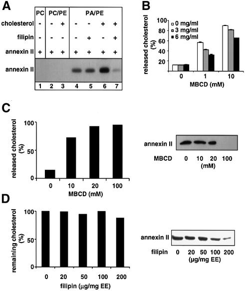
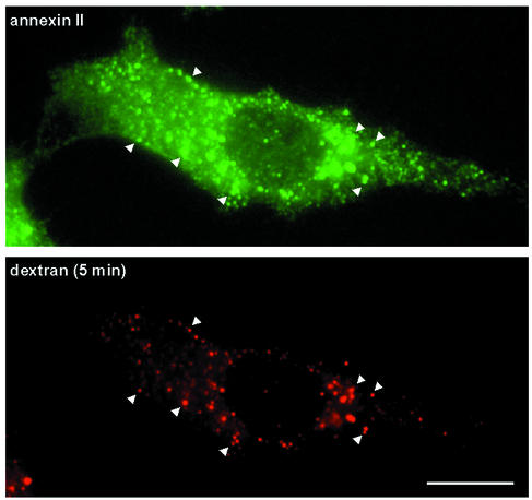
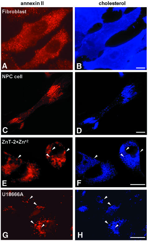
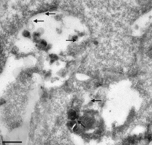
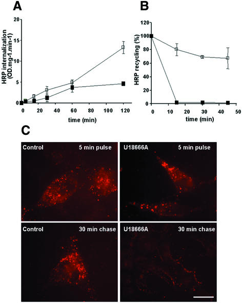
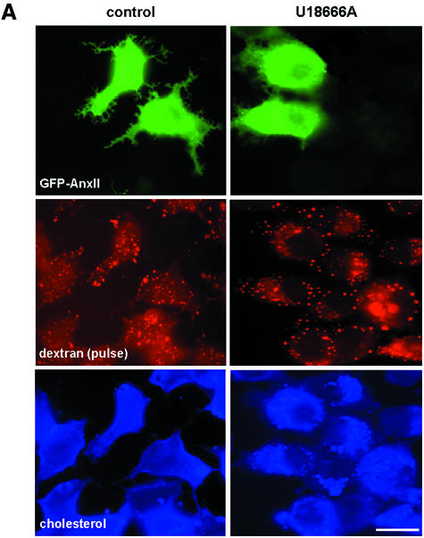
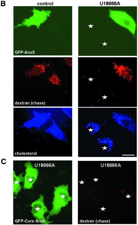
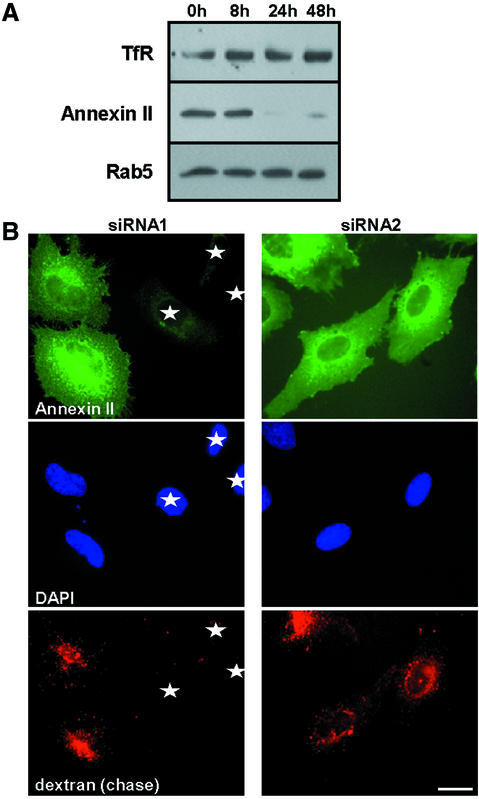
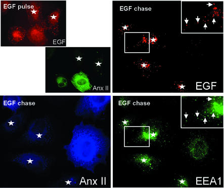
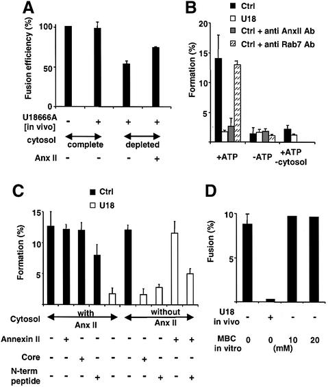
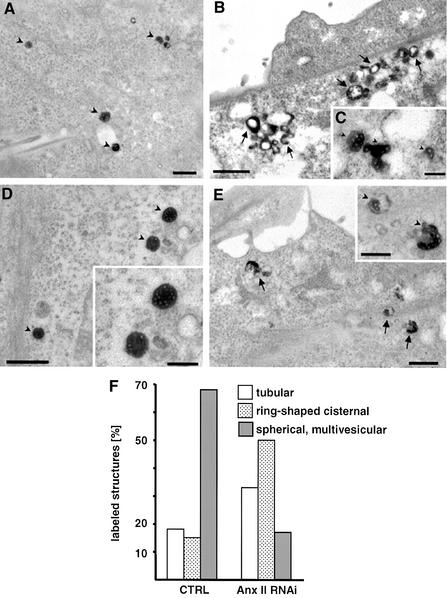
Proteins of the annexin family are believed to be involved in membrane-related processes, but their precise functions remain unclear. Here, we have made use of several experimental approaches, including pathological conditions, RNA interference and in vitro transport assays, to study the function of annexin II in the endocytic pathway. We find that annexin II is required for the biogenesis of multivesicular transport intermediates destined for late endosomes, by regulating budding from early endosomes-but not the membrane invagination process. Hence, the protein appears to be a necessary component of the machinery controlling endosomal membrane dynamics and multivesicular endosome biogenesis. We also find that annexin II interacts with cholesterol and that its subcellular distribution is modulated by the subcellular distribution of cholesterol, including in cells from patients with the cholesterol-storage disorder Niemann-Pick C. We conclude that annexin II forms cholesterol-containing platforms on early endosomal membranes, and that these platforms regulate the onset of the degradation pathway in animal cells.


Figures

None
Fig. 1. Binding to liposomes and early endosomes. (A) Liposomes of the following composition were prepared: PC only, PC/PE (1:1), PC/PE/cholesterol (2:2:1), PA/PE (1:1), PA/PE/cholesterol (2:2:1), and incubated as indicated with 1 µg of purified heterotetrameric annexin II and 5 mM EGTA for 30 min at room temperature. Lane 7 shows liposomes that were subsequently re-incubated with filipin for 30 min at 4°C. Liposomes were then collected by centrifugation and analyzed by SDS–PAGE (6 µmoles lipid/lane) and western blotting using the HH7 antibody against annexin II. (B) PA/PE/[1α-,2α(n)-3H] cholesterol (2:2:1) liposomes were incubated with the indicated annexin II concentrations (as in A), collected by floatation on gradients and reincubated at 4°C for 30 min with MBCD. Liposomes were collected as above and analyzed by liquid scintillation counting. Data are expressed as a percentage of the total cholesterol in each experiment (liposome and released). (C and D) Early endosomes were prepared from BHK cells labeled to equilibrium with [3H]cholesterol, incubated as in (A) with MBCD (C) or filipin [D; µg/mg early endosomal (EE) protein], and collected by centrifugation. Cholesterol release was quantified as in (B), and annexin analyzed as in (A); each lane contained 30 µg protein. Each experiment was repeated more than three times and representative experiments are shown.
None
Fig. 2. Annexin II in early endosomes. BHK cells were incubated for 5 min at 37°C with rhodamine-dextran to label early endosomes, permeabilized and fixed as described in Materials and methods, and then labeled with the monoclonal HH7 antibody against annexin II. Bar: 5 µm.
None
Fig. 3. Redistribution to late endosomes loaded with cholesterol. (A and B) Human skin fibroblasts from healthy individuals were labeled with filipin or anti-annexin II antibodies Annexin II was labeled with rhodamine-labeled secondary antibodies and filipin was directly visualized in the UV light range. (C and D) Human NPC skin fibroblasts double-labeled with filipin and anti-annexin II antibodies were analyzed as in (A and B). (E and F) Cholesterol accumulation was induced in BHK cells expressing the Zn2+ transporter ZnT-2 by incubation for 72 h with 0.1 mM ZnCl2 (Palmiter et al., 1996; Kobayashi et al., 1999). Cells were then processed as in (A–D). (G and H) BHK cells were incubated for 16 h with 3 µg/ml U18666A, and processed as above. Bars: 5 µm.
None
Fig. 4. Ultrastructural analysis of annexin II distribution in late endosomes loaded with cholesterol. BHK cells were incubated with U18666A as above, and processed for cryo-sectioning. Frozen sections were labeled with the HH7 anti-annexin II antibody followed by 10 nm goat anti-mouse gold particles (arrows). Bar: 0.1 µm.
None
Fig. 5. Internalization, recycling and intracellular accumulation. (A) BHK cells pretreated (closed symbol) or not (open symbol) with U18666A, as in Figure 3G and H, were incubated with 3 mg/ml HRP at 37°C, for the indicated time periods. Amounts of HRP accumulated in cells were quantified (OD U/min/mg cellular protein) at each time point. (B) BHK cells treated as in (A) were incubated for 5 min at 37°C with 0.5 mg/ml HRP, washed and then reincubated for the indicated time in marker-free medium. At each time point, HRP was quantified in cells and in the medium. The figure shows HRP that remained cell associated, expressed as a percentage of the total (cell associated and regurgitated) HRP. Each panel shows the mean of three series of experiments. (C) BHK cells treated as in (A) were incubated for 5 min at 37°C with 3 mg/ml rhodamine-dextran (pulse), to label early endosomes, or subsequently reincubated for 30 min in the absence of the marker (chase). Cells were then processed for fluorescence microscopy. Bar: 5 µm.
None
Fig. 6. Transport to late endosomes after cholesterol accumulation. (A and B) BHK cells transfected with a plasmid encoding for GFP–annexin II (GFP–AnxII) were pretreated or not with U18666A, and incubated for 5 min at 37°C with 3 mg/ml rhodamine-dextran (pulse), to label early endosomes (A), or reincubated for 40 min in the absence of the marker to label late endosomes (chase) (B). Cells were then processed as in Figure 3 and analyzed by triple-channel fluorescence. Stars show non-transfected cells. (C) Cells were treated as in (B), except that they were transfected with the GFP-tagged core domain of annexin II; the distribution of dextran and the GFP–core is shown. Bar: 5 µm.
None
Fig. 7. Annexin II down-modulation inhibits solute transport to late endosomes. (A) HeLa cells were transfected for the indicated time with siRNA1, and analyzed by SDS–PAGE and western blotting, using antibodies against annexin II, the transferrin receptor (TfR) and Rab5. (B) HeLa cells were transfected for 24 h with siRNA1 or siRNA2; 3 mg/ml rhodamine-dextran was then endocytosed for 5 min at 37°C and chased for 40 min. Cells were then processed for fluorescence microscopy. Stars show cells containing undetectable amounts of both annexin II and dextran. The extent of annexin II down-regulation was compatible with (A), but panels show (rare) examples of cells still expressing annexin II for comparison. Annexin II association to endosomes is not clearly visible with the fixation/permeabilization protocol used to detect endocytosed dextran—in contrast to Figure 2. Bar: 5 µm.
None
Fig. 8. EGF-receptor down-regulation. HeLa cells were transfected for 24 h with siRNA1 (Figure 7), and incubated for a 5 min pulse at 37°C with 0.4 µg/ml biotin–EGF and phycoerythrin-streptavidin, followed or not by a 60 min chase without the marker. Cells were then processed for immunofluorescence microscopy using anti-EEA1 antibodies and FITC-labeled secondary antibodies (EGF chase), as well as the HH7 anti-annexin II antibody and AMCA-labeled secondary antibodies, and analyzed by triple-channel immunofluorescence. As in Figure 7, annexin II association to endosomes is not clearly visible with this fixation/permeabilization protocol. Stars show cells containing undetectable amounts of annexin II. Bar: 5 µm.
None
Fig. 9. Biochemical analysis of endosomal transport. (A) The homotypic fusion of early endosomes was measured using early endosomes prepared from cells treated or not with U18666A, and cytosol depleted or not of annexin II. When indicated purified annexin II (AnxII) was added to the assay. Fusion is expressed as a percentage of the untreated control in complete cytosol. (B) Formation of ECV/MVBs was measured from donor early endosomal membranes prepared from cells treated (U18) or not (Ctrl) with U18666A, and using HRP as a marker of the endosomal content. In the assay, complete cytosol was supplemented or not with annexin II (AnxII) or antibodies (Ab) against annexin II or Rab7. Amounts of HRP sequestered within ECVs formed in vitro corresponded to ∼12% of the total early endosomal content, as expected (Aniento et al., 1993). (C) Formation of ECV/MVBs in vitro was as in (B), but the cytosol was depleted or not of annexin II and supplemented or not with purified annexin II, the core C-terminal domain of annexin II or the N-terminal peptide. (D) Donor membranes were prepared from cells treated or not with U18666A in vivo, as in (B), and then treated or not with MBCD at the indicated concentrations in vitro. After separation on gradients, the fusion capacity of these ECV/MVBS with late endosomes was measured in vitro.
None
Fig. 10. Endosome ultrastructure after annexin II down-regulation. In siRNA1- or mock-treated HeLa cells, microtubules were depolymerized with 10 µM nocodazole for 2 h, and HRP endocytosed for 5 min followed by a 45 min chase. (AE) In control cells (A and D), HRP labels predominantly ECV/MVBs (arrowheads) without associated tubules. In siRNA1-treated cells (B, C and E), HRP was mainly concentrated within ring-shaped and tubular structures with the typical morphology of early endosomes (arrows). Some multivesicular structures were labeled (C) but they lacked the uniform shape of the ECVs in the mock-treated cells (compare insets in D and E). (F) In 10 cell profiles chosen at random, HRP-labeled structures were classified according to morphology: tubular, thin tubules without associated multivesicular regions; ring-shaped cisternal, ring-shaped structures surrounding an electron-lucent space sometimes including multivesicular areas—resemble early endosomes; and spherical multivesicular, ECV/MVB. Each category is expressed as a percentage of the total number of labeled structures. Bars (A and B): 1 µm; (C and inset): 0.5µm.

Similar articles

Cited by

References

    1. Aniento F., Emans,N., Griffiths,G. and Gruenberg,J. (1993) Cytoplasmic dynein-dependent vesicular transport from early to late endosomes. J. Cell Biol., 123, 1373–1387. - PMC - PubMed
    1. Aniento F., Gu,F., Parton,R.G. and Gruenberg,J. (1996) An endosomal bCOP is involed in the pH-dependent formation of transport vesicles destined for late endosomes. J. Cell Biol., 133, 29–41. - PMC - PubMed
    1. Babiychuk E.B. and Draeger,A. (2000) Annexins in cell membrane dynamics. Ca(2+)-regulated association of lipid microdomains. J. Cell Biol., 150, 1113–1124. - PMC - PubMed
    1. Bishop N., Horman,A. and Woodman,P. (2002) Mammalian class E vps proteins recognize ubiquitin and act in the removal of endosomal protein–ubiquitin conjugates. J. Cell Biol., 157, 91–101. - PMC - PubMed
    1. Blackwood R.A. and Ernst,J.D. (1990) Characterization of Ca2+-dependent phospholipid binding vesicle aggregation and membrane fusion by annexins. Biochem. J., 266, 195–200. - PMC - PubMed

Publication types