. 2003 Jul 1;22(13):3267-78.
doi: 10.1093/emboj/cdg329.

Structural basis of the interaction between RalA and Sec5, a subunit of the sec6/8 complex

Affiliations

Structural basis of the interaction between RalA and Sec5, a subunit of the sec6/8 complex

Shuya Fukai et al. EMBO J. .

Abstract

The sec6/8 complex or exocyst is an octameric protein complex that functions during cell polarization by regulating the site of exocytic vesicle docking to the plasma membrane, in concert with small GTP-binding proteins. The Sec5 subunit of the mammalian sec6/8 complex binds Ral in a GTP-dependent manner. Here we report the crystal structure of the complex between the Ral-binding domain of Sec5 and RalA bound to a non-hydrolyzable GTP analog (GppNHp) at 2.1 A resolution, providing the first structural insights into the mechanism and specificity of sec6/8 regulation. The Sec5 Ral-binding domain folds into an immunoglobulin-like beta-sandwich structure, which represents a novel fold for an effector of a GTP-binding protein. The interface between the two proteins involves a continuous antiparallel beta-sheet, similar to that found in other effector/G-protein complexes, such as Ras and Rap1A. Specific interactions unique to the RalA.Sec5 complex include Sec5 Thr11 and Arg27, and RalA Glu38, which we show are required for complex formation by isothermal titration calorimetry. Comparison of the structures of GppNHp- and GDP-bound RalA suggests a nucleotide-dependent switch mechanism for Sec5 binding.


Figures
None
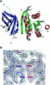
Fig. 1. Structure of the complex of the Sec5 Ral-binding domain and GppNHp-bound RalA and a representative σA-weighted 2FoFc electron density map. (A) Ribbon representation. The Sec5 Ral-binding domain consists of only β-strands and loops. The β-strands of the Sec5 Ral-binding domain are colored blue. The α-helices and β-strands of the GppNHp-bound RalA are colored red and green, respectively. The GppNHp molecule and the coordinated Mg2+ are shown as ball-and-stick models. For each protein, the α-helices and β-strands are numbered in sequential order. (B) The final refined model (shown as sticks) is superposed on the electron density map. The carbon atoms of the Sec5 Ral-binding domain and GppNHp-bound RalA are colored slate blue and pink, respectively. Water molecules are shown as balls colored cyan. The electron density map is contoured at the 1.3σ level around Sec5 Arg27 and RalA Glu38, showing a portion of the interface between the Sec5 Ral-binding domain and GppNHp-bound RalA.
None
Fig. 2. Comparison between the Sec5 Ral-binding domain and the NFκB p50 dimerization domain. (A) Structure of the NFκB p50 dimerization domain (PDB code 1IKN). (B) Structure of the Sec5 Ral-binding domain. (C) Schematic drawing of the topology of the Sec5 Ral-binding domain. β-strands are shown as blue arrows.
None
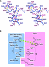
Fig. 3. The interaction between the Sec5 Ral-binding domain and GppNHp-bound RalA. (A) Stereo view of a stick representation. The carbon atoms of the Sec5 Ral-binding domain and GppNHp-bound RalA are colored slate blue and pink, respectively. Labels within slate blue and pink rectangles correspond to Sec5 and RalA residues, respectively. Hydrogen bonds are shown as dotted yellow lines. (B) Schematic drawing of the interaction between the Sec5 Ral-binding domain and GppNHp-bound RalA. Hydrogen bonds are shown as dotted yellow lines. A stacking interaction is shown as a dotted green bold line.
None
Fig. 4. Sequence alignments of the Ral-binding domain of Sec5 and the Sec5-interacting region of Ral. (A) Sequence alignment of the Ral-binding domain of Sec5. The secondary structure is shown at the top. The amino acid residues whose side chains interact with RalA are highlighted with cyan rectangles. (B) Sequence alignment of the Sec5-interacting region of RalA. The secondary structure is shown at the top. The RalA amino acid residues whose side chains interact with the Sec5 Ral-binding domain are highlighted with pink rectangles. The corresponding amino acid residues in Ras/Rap are highlighted with orange rectangles.
None
Fig. 5. Comparison of the interface between the Sec5 Ral-binding domain and GppNHp-bound RalA with that between GppNHp-bound Ras/Rap and their corresponding Ras binding domains (RBDs). A ribbon representation is shown at the top of each panel. The α-helices and β-strands of the Sec5 Ral-binding domain/RBDs are colored brown and blue, respectively, while those of GppNHp-bound Ral/Ras/Rap are colored red and green, respectively. The GppNHp molecule and the coordinated Mg2+ are shown as ball-and-stick models. At the bottom of each panel, the amino acid residues involved in the intermolecular β-sheet are shown as sticks in two orientations differing by 90°. The carbon atoms of the Sec5 Ral-binding domain/RBDs and the GppNHp-bound Ral/Ras/Rap are colored slate blue and pink, respectively. The view angle of the left diagram is nearly the same as that of the ribbon representation drawn at the top of each panel.
None
Fig. 6. Isothermal calorimetric titration of RalA with the Sec5 Ral-binding domain. The top panels show raw data obtained after 5 µl injections of 150 µM Sec5 RalA-binding domain into 4 µM RalA in 50 mM PBS containing 5 mM MgCl2 and 0.5 mM GppNHp or GDP. The bottom panels show a non-linear least-squares fit (–) of the heat released as a function of the added ligand. (A) RalA titration with Sec5 in the presence of GppNHp. (B) RalA titration with Sec5 in the presence of GDP. (C) RalA (Glu38Ala) titration with Sec5 in the presence of GppNHp. Bottom panel: solid circles for RalA (Glu38Ala) and open circles for RalA (Glu38Arg). (D) RalA titration with 110 µM Sec5 (Thr11Ala) in the presence of GppNHp. Bottom panel: solid diamonds for Sec5 (Thr11Ala) titration and open diamonds for Sec5 (Arg27Ala) titration. Note that the raw data for RalA (Glu38Arg)–Sec5 and RalA–Sec5 (Arg27Ala) in the presence of GppNHp were similar to that seen for RalA–Sec5 in the presence of GDP.
None
Fig. 7. Guanine-nucleotide-dependent conformational change in RalA. (A) The GDP-bound RalA is superposed on the GppNHp-bound RalA in complex with the Sec5 Ral-binding domain. A ribbon representation is shown. The Sec5 Ral-binding domain is colored in green. The GDP- and GppNHp-bound RalA molecules are colored in white, except for the switch I (residues 40–48) and II (residues 70–78) regions. The coloring schemes for switch I and II are described in the figure. (B) Interactions of the γ-phosphate of GppNHp with switch I and II (stereo view). The amino acid residues around the γ-phosphate are shown as sticks. The carbon atoms of switch I, switch II and the other regions of RalA are colored in slate blue, orange and white, respectively. The carbon and phosphorus atoms of the GppNHp molecule are colored in green and yellow, respectively. The coordinated Mg2+ and water molecules are shown as gray and cyan balls, respectively. Dotted yellow lines indicate hydrogen bonds.

Similar articles

Cited by

References

    1. Abrahams J.P. and Leslie,A.G.W. (1996) Methods used in the structure determination of bovine mitochondrial F1 ATPase. Acta Crystallogr. D, 52, 30–42. - PubMed
    1. Adamo J.E., Rossi,G. and Brennwald,P. (1999) The Rho GTPase Rho3 has a direct role in exocytosis that is distinct from its role in actin polarity. Mol. Biol. Cell, 10, 4121–4133. - PMC - PubMed
    1. Adams P.D., Pannu,N.S., Read,R.J. and Brunger,A.T. (1997) Cross-validated maximum likelihood enhances crystallographic simulated annealing refinement. Proc. Natl Acad. Sci. USA, 94, 5018–5023. - PMC - PubMed
    1. Altschul S.F., Gish,W., Miller,W., Myers,E.W. and Lipman,D.J. (1990) Basic local alignment search tool. J. Mol. Biol., 215, 403–410. - PubMed
    1. Altschul S.F., Madden,T.L., Schaffer,A.A., Zhang,J., Zhang,Z., Miller,W. and Lipman,D.J. (1997) Gapped BLAST and PSI-BLAST: a new generation of protein database search programs. Nucleic Acids Res., 25, 3389–3402. - PMC - PubMed

Publication types