. 2003 Jul 1;22(13):3279-90.
doi: 10.1093/emboj/cdg317.

A direct role for GRASP65 as a mitotically regulated Golgi stacking factor

Affiliations

A direct role for GRASP65 as a mitotically regulated Golgi stacking factor

Yanzhuang Wang et al. EMBO J. .

Erratum in

  • EMBO J. 2003 Aug 1;22(15):4026

Abstract

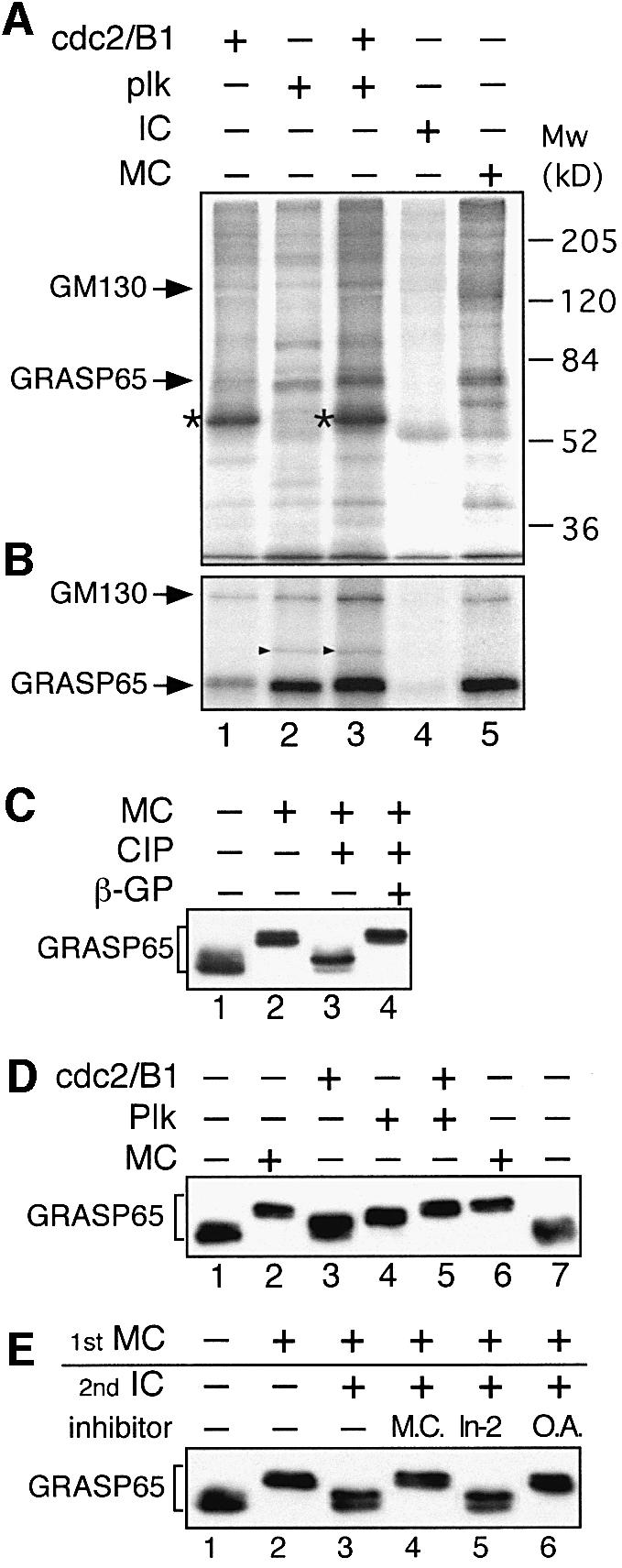
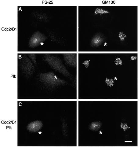
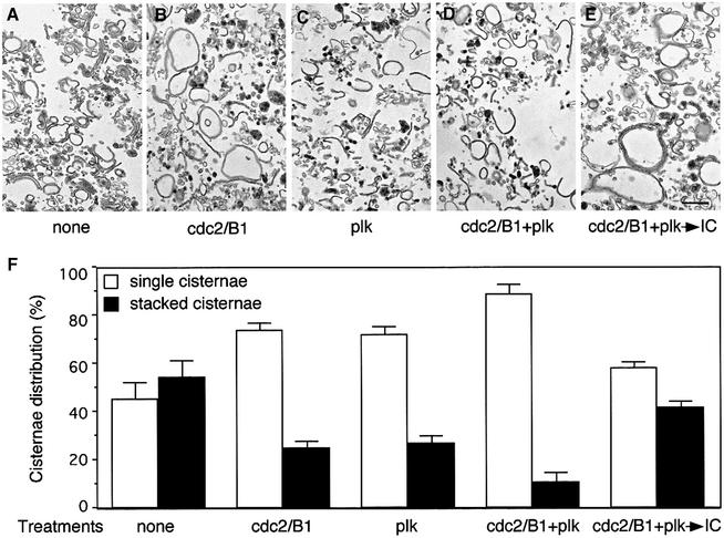
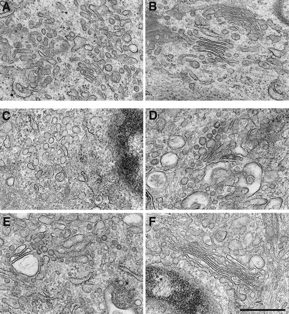
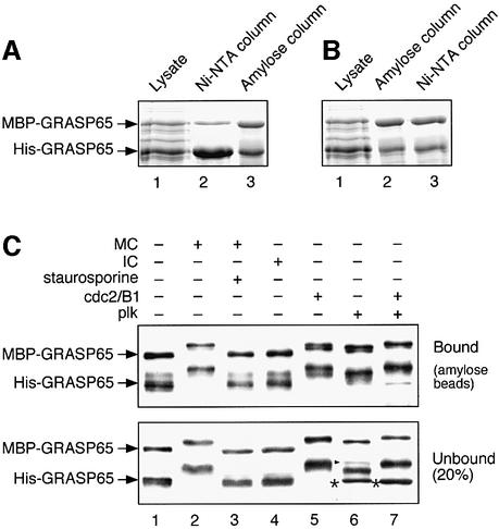
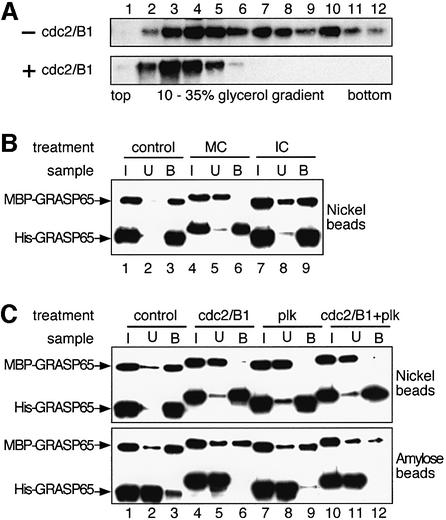
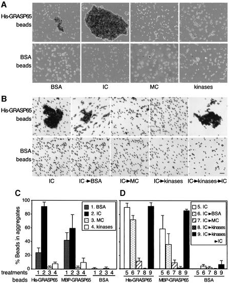
Cell-free assays that mimic the disassembly and reassembly cycle of the Golgi apparatus during mitosis implicated GRASP65 as a mitotically regulated stacking factor. We now present evidence that GRASP65 is directly involved in stacking Golgi cisternae. GRASP65 is the major phosphorylation target in rat liver Golgi membranes of two mitotic kinases, cdc2-cyclin B and polo-like kinases, which alone will unstack Golgi membranes, generating single cisternae. Mitotic cells microinjected with antibodies to GRASP65 fail to form proper Golgi stacks after cell division. Beads coated with GRASP65 homodimers form extensive aggregates consistent with the formation of trans oligomers. These can be disaggregated using purified cdc2-cyclin B1 and polo-like kinases, and re-aggregated after dephosphorylation of GRASP65. Together, these data demonstrate that GRASP65 has the properties required to bind surfaces together in a mitotically regulated manner.


Figures

None
Fig. 1. GRASP65 is the major target of cdc2/B1 and plk kinases on Golgi membranes. (A) Membranes were incubated in the presence of [γ-32P]ATP with purified cdc2/B1 (lane 1), plk (lane 2), cdc2/B1 and plk (lane 3), interphase cytosol (IC, lane 4) or mitotic cytosol (MC, lane 5). Kinases and MC were adjusted to equivalent levels of kinase activity. The membranes were isolated and analyzed by SDS–PAGE followed by autoradiography. Arrows indicate phosphorylated GM130 and GRASP65. The strongly labeled protein in lanes 1 and 3 (asterisks) is cyclin B1. (B) Membrane samples from (A) were solubilized and GRASP65 immunoprecipitated followed by SDS–PAGE and autoradiography. Phosphorylated GM130 co-precipitated with phosphorylated GRASP65. The additional band in lanes 2 and 3 is GST–plk (arrowheads) which co-precipitated with GRASP65. (C) Detection of phosphorylated GRASP65 by a bandshift assay. RLG membranes were treated with either buffer (lane 1) or MC (lanes 2–4). Membranes were isolated and treated with alkaline phosphatase (CIP) in the absence (lane 3) or presence (lane 4) of β-glycerophosphate (β-GP), and analyzed by immunoblotting. (D) RLG membranes were incubated with the indicated kinases and immunoblotted for GRASP65. Note that the combination of cdc2/B1 and plk kinases phosphorylated GRASP65 to a similar extent as MC. (E) Phosphorylation and dephosphorylation of GRASP65 by MC and IC. RLG membranes were first incubated with MC (lanes 2–6) and then IC (lanes 3–6), in the absence or presence of microcystin (M.C., lane 4), Inhibitor-2 (In-2, lane 5) or okadaic acid (O.A., lane 6), followed by immunoblotting for GRASP65. Note that microcystin and okadaic acid inhibited dephosphorylation by IC, implicating PP2A in this process.
None
Fig. 2. Microinjection of cdc2/B1 but not plk kinases leads to GM130 phosphorylation and Golgi fragmentation. NRK cells were injected with cdc2/B1 (A), plk (B) or cdc2/B1 and plk (C). Biotinylated BSA was used as an injection marker (asterisks). After 30 min at 37°C, cells were fixed and double labeled with polyclonal antibodies to phosphorylated GM130 (PS-25, left panels) and a monoclonal antibody to GM130 (GM130, right panels). Bar, 10 µm.
None
Fig. 3. Purified cdc2/B1 and plk kinases unstack Golgi cisternae. (AE) RLG stacks were either left untreated (A), or treated with cdc2/B1 (B), plk (C) or both (D) at 37°C for 20 min. Membranes were re-isolated after kinase treatment (D) and further treated with IC for 60 min at 37°C (E). Membranes were fixed and processed for EM. Bar, 0.5 µm. (F) Quantitation of (A–E), by the intersection method, to estimate the percentage of single or stacked cisternal membranes. Results represent the mean of three independent experiments ±SEM. Note the increased number of single cisternae after kinase treatments.
None
Fig. 4. Antibodies to GRASP65 prevent proper re-stacking of Golgi membranes in post-mitotic cells. NRK cells in metaphase were microinjected with affinity-purified, anti-GRASP65 antibodies (A and CE) or non-specific rabbit IgGs as a control (B and F). After completion of cell division, the injected cells were processed for EM. Results of two individual experiments are shown in (A and B) and (C–F), respectively. Note the properly assembled stacks in cells injected with IgGs (B and F) and the disorganized Golgi structures in cells injected with antibodies to GRASP65 (A and C–E). Bar, 0.5 µm.
None
Fig. 5. Recombinant GRASP65 forms stable dimers. (A and B) MBP-tagged GRASP65 and His-tagged GRASP65 were co-expressed in E.coli and purified sequentially on nickel followed by amylose columns (A), or the reverse (B). Samples were fractionated by SDS–PAGE. Note that the ratio of the two tagged proteins in the complex was ∼1:1, irrespective of the order of purification. (C) Purified heterodimers of MBP–GRASP65/His-GRASP65 bound to amylose beads were treated with buffer alone (lane 1), MC ( lane 2), MC and the general kinase inhibitor staurosporine (lane 3), IC (lane 4), cdc2/B1 (lane 5), plk (lane 6) or both (lane 7). Beads (bound, upper panel) or supernatant (unbound, lower panel) were analyzed by immunoblotting for GRASP65. The extra bands in lanes 6 and 7 are cGST–plk (arrowhead) and its fragments (asterisk). Note that none of the treatments changed the ratio of the two proteins in the bound or unbound fractions.
None
Fig. 6. GRASP65 dimers form higher order oligomers in a cell cycle-dependent manner. (A) His-GRASP65 was incubated in the absence or presence of cdc2/B1, sedimented in glycerol gradients and fractions analyzed by western blotting. Note that the mixture of GRASP65 oligomers is reduced to a single oligomeric complex after treatment with mitotic kinase. (B) Separately expressed and purified MBP–GRASP65 and His-GRASP65 proteins were mixed and incubated in the presence of buffer (control, lanes 1–3), MC (lanes 4–6) or IC (lanes 7–9). The protein complex was then isolated using nickel beads. Equal portions of the input (I), unbound (U) or bound (B) fraction were analyzed by immunoblotting for GRASP65. Note the absence of MBP–GRASP65 in the bound complexes treated with MC (lane 6). (C) As in (B) except that purified dimers were incubated with cdc2/B1 and plk alone or in combination. Complexes were isolated on nickel beads (upper panel) or amylose beads (lower panel) and processed as in (B). Note that bound complexes under control conditions contained both tagged GRASP65 proteins (lane 3) whereas kinase treatment separated them (lanes 6, 9 and 12).
None
Fig. 7. GRASP65 can form trans oligomers. (A) Purified His-GRASP65 (upper panel), or BSA (as control, lower panel) was covalently coupled to Dynal beads and incubated with BSA, IC, MC or cdc2/B1 and plk (kinases). After incubation, the beads were placed on glass slides and random fields photographed. A representative image of each condition is shown. Bar, 500 µm. (B) As in (A) except that the GRASP65 beads were first aggregated using IC and then treated with either BSA, MC or a combination of cdc2/B1 and plk (kinases). Kinase-treated beads were treated further with IC (IC→kinases→IC). Bar, 500 µm. (C and D) Quantitation of (A) and (B) and MBP–GRASP65 beads. Note that aggregates only formed in the presence of buffer and IC and that these aggregates were disassembled reversibly by MC and recombinant kinases.

Similar articles

Cited by

References

    1. Barr F.A., Puype,M., Vandekerckhove,J. and Warren,G. (1997) GRASP65, a protein involved in the stacking of Golgi cisternae. Cell, 91, 253–262. - PubMed
    1. Barr F.A., Nakamura,N. and Warren,G. (1998) Mapping the interaction between GRASP65 and GM130, components of a protein complex involved in the stacking of Golgi cisternae. EMBO J., 17, 3258–3268. - PMC - PubMed
    1. Barr F.A., Preisinger,C., Kopajtich,R. and Korner,R. (2001) Golgi matrix proteins interact with p24 cargo receptors and aid their efficient retention in the Golgi apparatus. J. Cell Biol., 155, 885–891. - PMC - PubMed
    1. Dirac-Svejstrup A.B., Shorter,J., Waters,M.G. and Warren,G. (2000) Phosphorylation of the vesicle-tethering protein p115 by a casein kinase II-like enzyme is required for Golgi reassembly from isolated mitotic fragments. J. Cell Biol., 150, 475–488. - PMC - PubMed
    1. Draviam V.M., Orrechia,S., Lowe,M., Pardi,R. and Pines,J. (2001) The localization of human cyclins B1 and B2 determines CDK1 substrate specificity and neither enzyme requires MEK to disassemble the Golgi apparatus. J. Cell Biol., 152, 945–958. - PMC - PubMed

Publication types