. 2003 Jul 1;22(13):3291-302.
doi: 10.1093/emboj/cdg325.

Structure and axon outgrowth inhibitor binding of the Nogo-66 receptor and related proteins

Affiliations

Structure and axon outgrowth inhibitor binding of the Nogo-66 receptor and related proteins

William A Barton et al. EMBO J. .

Abstract

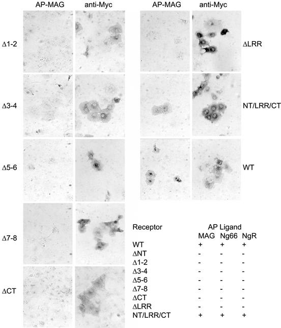
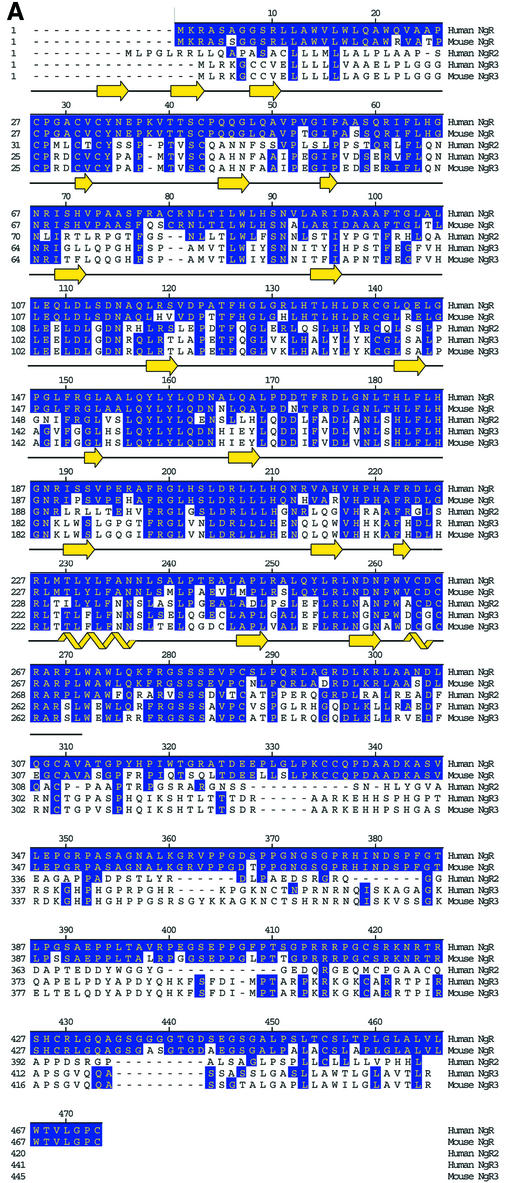
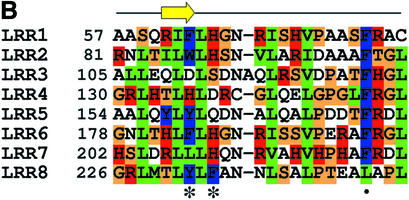
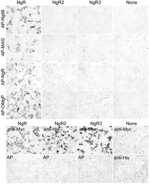
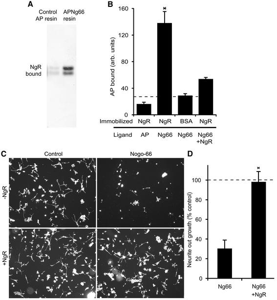
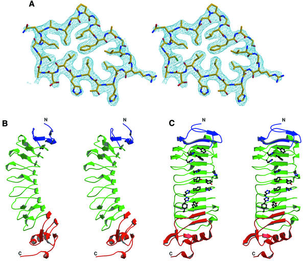
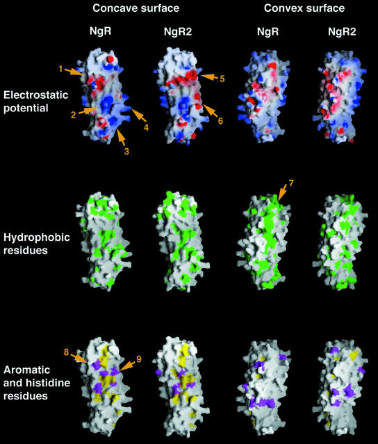
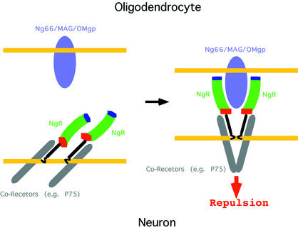
The myelin-derived proteins Nogo, MAG and OMgp limit axonal regeneration after injury of the spinal cord and brain. These cell-surface proteins signal through multi-subunit neuronal receptors that contain a common ligand-binding glycosylphosphatidylinositol-anchored subunit termed the Nogo-66 receptor (NgR). By deletion analysis, we show that the binding of soluble fragments of Nogo, MAG and NgR to cell-surface NgR requires the entire leucine-rich repeat (LRR) region of NgR, but not other portions of the protein. Despite sharing extensive sequence similarity with NgR, two related proteins, NgR2 and NgR3, which we have identified, do not bind Nogo, MAG, OMgp or NgR. To investigate NgR specificity and multi-ligand binding, we determined the crystal structure of the biologically active ligand-binding soluble ectodomain of NgR. The molecule is banana shaped with elongation and curvature arising from eight LRRs flanked by an N-terminal cap and a small C-terminal subdomain. The NgR structure analysis, as well as a comparison of NgR surface residues not conserved in NgR2 and NgR3, identifies potential protein interaction sites important in the assembly of a functional signaling complex.


Figures

None
Fig. 1. MAG-binding region in NgR. COS-7 cells were transfected with wild-type (WT) NgR or NgR deletion mutant plasmids and tested for AP-MAG binding. WT NgR and LRRNT/LRR/LRRCT-expressing COS-7 cells bind AP-MAG, whereas other NgR deletion mutants do not. Table I summarizes the ligand-binding attributes of the different NgR mutants from the present work and from Fournier et al. (2002). Deletions were as follows: NgR-ΔNT, residues 27–57; NgR-Δ1–2, residues 58–105; NgR-Δ3–4, residues 106–154; NgR-Δ5–6, residues 155–202; NgR-Δ7–8, residues 203–250; NgR-ΔCT, residues 260–310; NgR-ΔLRR, residues 27–310; and NT/LRR/CT, residues 311–445.
None
Fig. 2. Comparison of NgR, NgR2 and NgR3 protein sequence. (A) Alignment of the amino acid sequence of the human and mouse NgR, human NgR2 and human and mouse NgR3. Regions of sequence identity are indicated. Secondary structure present in the NgR (27–311) crystal structure is indicated. The black line indicates the extent of the crystallographic model. (B) Sequence alignment of the individual NgR repeats that constitute the LRR domain (Fournier et al., 2001). Conserved structurally important residues are in green, and the residues with exposed aromatic and polar side chains are in blue and red, respectively. Asterisks indicate the positions of the aromatic and histidine exposed residues highlighted in Figure 6. The dot denotes the position of the conserved phenylalanine present in ‘typical’ LRRs.
None
Fig. 3. NgR2 and NgR3 do not bind NgR ligands. COS-7 cells were transfected with vectors for Myc-NgR, His-NgR2, Myc-NgR3 or None and then stained with AP-tagged soluble ligands as indicated (upper half). The AP-ligand concentration was 10 nM AP-Nogo-66, 20 nM AP-MAG, 20 nM AP-NgR and 20 nM AP-OMgp. Note the expression of NgR2 and NgR3 without detectable Nogo-66, MAG, NgR or OMgp binding. In the lower half, the expression of recombinant epitope-tagged NgR, NgR2 and NgR3 proteins is detected by immunostaining with the indicated anti-epitope-tag antibodies, anti-Myc or anti-His. None of the proteins binds 20 nM AP protein (bottom row).
None
Fig. 4. Biological activity of the purified recombinant NgR protein. (A) Control AP protein or AP-Nogo-66 were bound to resin and then incubated with NgR. After washing, bound protein was examined by NgR immunoblot. The two bands on the gel correspond to differently glycosylated forms of NgR. (B) Microtiter wells were coated with the indicated proteins and then probed with AP-Nogo-66 or AP protein in the presence or absence of excess soluble NgR. (C) Rat P4-6 DRG neurons were plated on surfaces coated with or without GST–Nogo-66 and with or without the addition of excess NgR as indicated. Rhodamine-phalloidin staining is illustrated. (D) Neurite outgrowth from an experiment as in (C) is reported as a percentage of the value without GST–Nogo-66. Data are means ± SEM for three or more measurements. *Values with NgR are significantly different from those without NgR (p ≤ 0.02, Student’s t-test). Dissociated rat P4-6 DRG neurons were plated on surfaces coated with 90 ng of control GST or 90 ng of GST–Nogo-66 with the addition of 450 ng of NgR protein or 450 ng of control GST protein. After 6 h, cells were fixed and scored for neurite outgrowth per neuron as described previously (Fournier et al., 2002; Liu et al., 2002).
None
Fig. 5. Structure of NgR. (A) Representative region of the density-modified experimental electron density map contoured at 1.5 σ. The refined NgR model is shown with carbons in yellow, nitrogens in blue and oxygens in red. (B and C) Orthogonal stereoviews of the NgR structure, the LRRNT subdomain is in blue, the central LRR subdomain in green and the LRRCT subdomain in red. The protein N- and C-termini are indicated. The side chains of the exposed aromatic and histidine residues lining the concave molecular surface are shown only in (C).
None
Fig. 6. The molecular surface of NgR and NgR2 colored according to electrostatic potential (top row), hydrophobicity (middle row) or with colored exposed aromatic residues in yellow and histidines in magenta (bottom row). The model for the NgR2 structure was generated using the known structure of NgR (the two proteins share a 60% aa identity in the modeled region). Important surface regions, discussed in the text, are indicated with numbered arrows. The molecules are positioned with their N-termini pointing up and their C-termini pointing down.
None
Fig. 7. A model for initiation of the repulsive signaling mediated by NgR. Although recombinant NgR (NT/LRR/CT) is monomeric in solution, it has been shown to associate at the cell surface with itself (Fournier et al., 2002) and with its co-receptor p75 (Wang et al., 2002b). NgR interacts with a variety of structurally unrelated ligands, some of which may bind with 2:2 and some with 2:1 stoichiometry.

Similar articles

Cited by

References

    1. Bandtlow C., Zachleder,T. and Schwab,M.E. (1990) Oligodendrocytes arrest neurite growth by contact inhibition. J. Neurosci., 10, 3837–3848. - PMC - PubMed
    1. Bartsch U. et al. (1995) Lack of evidence that myelin-associated glycoprotein is a major inhibitor of axonal regeneration in the CNS. Neuron, 15, 1375–1381. - PubMed
    1. Brunger A.T. et al. (1998) Crystallography & NMR system: a new software suite for macromolecular structure determination. Acta Crystallogr. D, 54, 905–921. - PubMed
    1. CCP4 (1994) The CCP4 suite: programs for X-ray crystallography. Acta Crystallogr. D, 50, 760–763. - PubMed
    1. David S. and Aguayo,A.J. (1981) Axonal elongation into peripheral nervous system ‘bridges’ after central nervous system injury in adult rats. Science, 214, 931–933. - PubMed

Publication types