. 2003 Jul 1;22(13):3303-13.
doi: 10.1093/emboj/cdg328.

A role for maternal beta-catenin in early mesoderm induction in Xenopus

Affiliations

A role for maternal beta-catenin in early mesoderm induction in Xenopus

Anne Schohl et al. EMBO J. .

Abstract

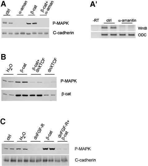
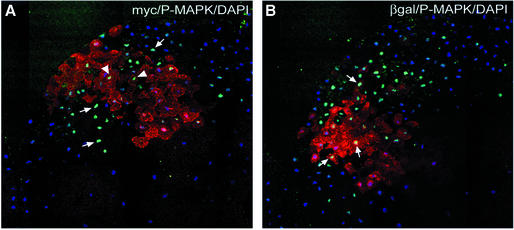
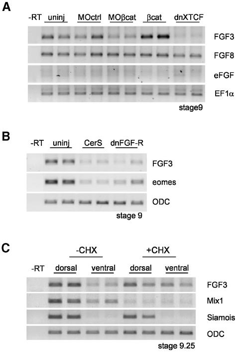
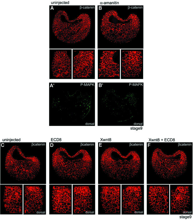
Mesoderm formation results from an inducing process that requires maternal and zygotic FGF/MAPK and TGFbeta activities, while maternal activation of the Wnt/beta-catenin pathway determines the anterior-dorsal axis. Here, we show a new role of Wnt/beta-catenin signaling in mesoderm induction. We find that maternal beta-catenin signaling is not only active dorsally but also all around the equatorial region, coinciding with the prospective mesoderm. Maternal beta-catenin function is required both for expression of dorsal genes and for activation of MAPK and the mesodermal markers Xbra and eomesodermin. beta-catenin acts in a non- cell-autonomous manner upstream of zygotic FGF and nodal signals. The Wnt/beta-catenin activity in the equatorial region of the early embryo is the first example of a maternally provided mesoderm inducer restricted to the prospective mesoderm.


Figures

None
Fig. 1. β-catenin is required for activation of MAPK and early expression of pan-mesodermal genes. Embryos were injected four times equatorially at the four-cell stage and fixed or extracted at the indicated stages. (A and B) Double staining for β-catenin (A and B) and P-MAPK (A′ and B′) of cryosections from embryos injected with (A) control morpholino oligonucleotides (MO control, 4 × 5 ng) or (B) β-catenin MO (MO β-catenin, 4 × 5 ng). Small panels show details of β-catenin staining in the ventral and dorsal marginal zone. Brackets indicate marginal zone; arrows point to β-catenin and P-MAPK positive nuclei; an, animal pole; vg, vegetal pole; β-catenin MO depleted nuclear β-catenin on both the dorsal and the ventral side and reduced MAPK activation along the marginal zone. (C) Immunoblot of total extracts from embryos injected with control or β-catenin MO (4 × 5 ng) stained with anti-P-MAPK antibody or with anti-C-cadherin antibody used as loading control. β-catenin MO decreased the levels of activated P-MAPK. (D) RT–PCR analysis of pan-mesodermal (Xbra, eomesodermin), dorsal (gsc) and ventral (Wnt8) markers. Depletion of nuclear β-catenin (MOβcat, 4 × 5 ng) or downregulation of β-catenin signaling by injection of dominant-negative XTFC3 mRNA (dnXTCF, 4 × 2 ng) inhibited expression of the early mesodermal genes Xbra and eomesodermin and of gsc. β-catenin overexpression (4 × 2 ng mRNA) stimulated expression of Xbra, eomesodermin and gsc and inhibited expression of Wnt8. ODC was used as loading control. (E) dnXTCF inhibits Xbra and eomesodermin expression not only on the dorsal side, but also on the ventral side. Control embryos and embryos injected with dnXTCF (4 × 0.5 ng) were dissected into dorsal and ventral halves at stage 9.5 and analyzed by RT–PCR. Siamois, a dorsal marker directly dependent on β-catenin signaling, was used as control for proper dissection.
None
Fig. 2. Activation of MAPK by β-catenin is indirect and requires FGF signaling. P-MAPK immunoblots of embryo extracts, stage 9. Embryos were injected with β-catenin mRNA (4 × 2 ng), α-amanitin (4 × 50 pg), dominant-negative XTFC3 mRNA (dnXTCF, 4 × 2 ng) or dominant-negative FGF receptor (dnFGF-R, 4 × 2 ng). Control, uninjected; H2O, water injected. Both normal and β-catenin-induced P-MAPK activation were blocked by (A) α-amanitin, (B) dnXTCF and (C) dnFGF-R. β-catenin blot in (B): co-expression of dnXTCF does not affect the levels of β-catenin. (A′) RT–PCR showing complete block of transcription of the zygotic gene Wnt8 in embryos injected with α-amanitin.
None
Fig. 3. Activation of MAPK by β-catenin is non-cell autonomous. Stage 16-cell embryos were injected ventrally in one blastomere with myc-tagged β-catenin mRNA (500 pg) and in an adjacent blastomere with β-galactosidase mRNA (1 ng). Embryos were fixed at stage 9 and analyzed by immunofluorescence on equatorial sections. (A) Double staining for myc-β-catenin (anti-myc, red) and P-MAPK (green). (B) Double staining for β-galactosidase (red) and P-MAPK (green). (A) and (B) show consecutive sections. Nuclei were stained with DAPI (blue). P-MAPK signal was not only increased in β-catenin-overexpressing cells (arrowheads in A), but also in adjacent cells (arrows).
None
Fig. 4. Regulation of FGF3 by early β-catenin, Xnr and FGF signals. (A) FGF3 is a target of β-catenin. FGF3, FGF8 and eFGF were analyzed by RT–PCR at stage 9. FGF3 expression was inhibited by β-catenin morpholino oligonucleotides (MOβcat, 4 × 5 ng) and dominant-negative XTFC3 mRNA (dnXTCF, 4 × 2 ng mRNA) and activated by β-catenin overexpression (4 × 2 ng mRNA). FGF8 was unaffected. eFGF was barely detectable at this early stage. EF1α was used as loading control. (BFGF3 and eomesodermin expression at stage 9 were inhibited by cerberus short (CerS, 4 × 150 pg mRNA) and dominant-negative FGF receptor (dnFGF-R, 4 × 2 ng mRNA). (C) Early FGF3 expression is resistant to cycloheximide (CHX) treatment. Embryos were cut into dorsal and ventral halves at stage 7, and the halves were cultured in the presence or absence of 100 µg/ml CHX until stage 9.25. Siamois was used as a dorsal marker and Mix1 as positive control for the effectiveness of CHX.
None
Fig. 5. Rescue of MAPK activity and eomesodermin expression by FGF3 and Xnr1. (A) P-MAPK blot of stage 9.25 extracts: P-MAPK was decreased by dominant-negative XTFC3 mRNA (dnXTCF, 4 × 2 ng mRNA) but could be rescued by co-expression of FGF3 (4 × 10 pg mRNA). Control, uninjected; H2O, water injected. (B) RT–PCR: expression of eomesodermin at stage 9.25 was strongly inhibited by dnXTCF (4 × 2 ng mRNA). Co-expression of FGF3 (4 × 10 pg mRNA) and Xnr1 (4 × 5 pg mRNA) could rescue normal levels of eomesodermin. FGF3 alone had no effect. Partial rescue was observed with Xnr1 alone, which was completely blocked by dominant-negative FGF receptor (dnFGF-R, 4 × 2 ng mRNA).
None
Fig. 6. Nuclear β-catenin in the marginal zone is maternal and independent of Wnt ligands. (A and B) Double staining for β-catenin (A and B) and P-MAPK (A′ and B′) of cryosections from (A) control embryos or (B) embryos injected with α-amanitin (stage 9, 4 × 50 pg). Small panels show enlargements of β-catenin staining in the ventral and dorsal marginal zone. α-amanitin strongly reduced the P-MAPK signal but had no effect on nuclear β-catenin, neither on the dorsal nor on the ventral side. (CF) β-catenin staining for (C) control embryos or for embryos injected with mRNA coding for (D) the extracellular domain of Xenopus Frizzled 8 (ECD8, 4 × 500 pg), (E) Xwnt8 (4 × 10 pg) or (F) ECD8 and Xwnt8. Xwnt8 strongly increased nuclear β-catenin on the ventral side (E), and this increase was prevented by co-injection of ECD8 (F). However, ECD8 had no effect on the endogenous ventral and dorsal β-catenin signals (compare C and D).
None
Fig. 7. Model for early mesoderm induction. Maternal β-catenin activity appears to be pre localized along the equator. It is strongest and most widespread in the dorsal side, where it induces dorsal genes, such as Siamois. Both β-catenin and VegT induce Xnrs. FGF, Xnr and β-catenin signals cooperate to induce FGF3 and, perhaps, other FGFs along the marginal zone and thus activate MAPK. The combination of Xnr and FGF/MAPK signals then activates expression of mesodermal genes such as eomesodermin and Xbra.

Similar articles

Cited by

References

    1. Agius E., Oelgeschlager,M., Wessely,O., Kemp,C. and De Robertis,E.M. (2000) Endodermal Nodal-related signals and mesoderm induction in Xenopus. Development, 127, 1173–1183. - PMC - PubMed
    1. Amaya E., Musci,T.J. and Kirschner,M.W. (1991) Expression of a dominant negative mutant of the FGF receptor disrupts mesoderm formation in Xenopus embryos. Cell, 66, 257–270. - PubMed
    1. Angerer L.M. and Angerer,R.C. (2000) Animal–vegetal axis patterning mechanisms in the early sea urchin embryo. Dev. Biol., 218, 1–12. - PubMed
    1. Cadigan K. and Nusse,R. (1997) Wnt signaling: a common theme in animal development. Genes Dev., 11, 3286–3305. - PubMed
    1. Campione M. et al. (1999) The homeobox gene Pitx2: mediator of asymmetric left–right signaling in vertebrate heart and gut looping. Development, 126, 1225–1234. - PubMed

MeSH terms