. 2003 Jul 1;22(13):3314-25.
doi: 10.1093/emboj/cdg335.

Arabidopsis AXR6 encodes CUL1 implicating SCF E3 ligases in auxin regulation of embryogenesis

Affiliations

Arabidopsis AXR6 encodes CUL1 implicating SCF E3 ligases in auxin regulation of embryogenesis

Hanjo Hellmann et al. EMBO J. .

Abstract

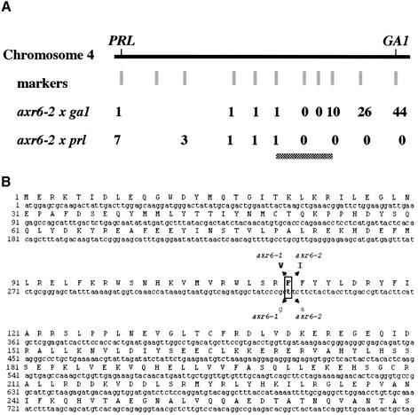
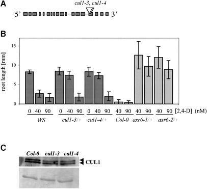
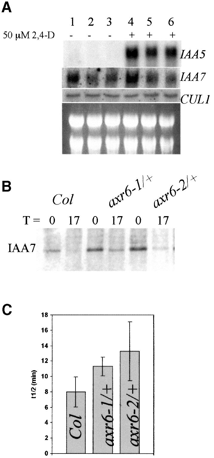
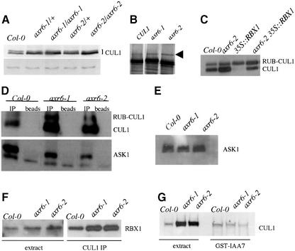
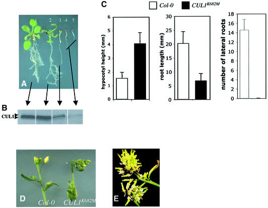
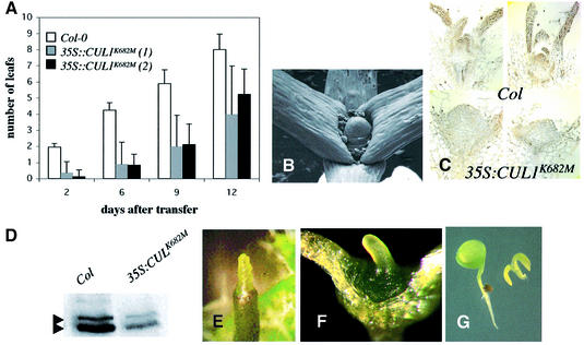
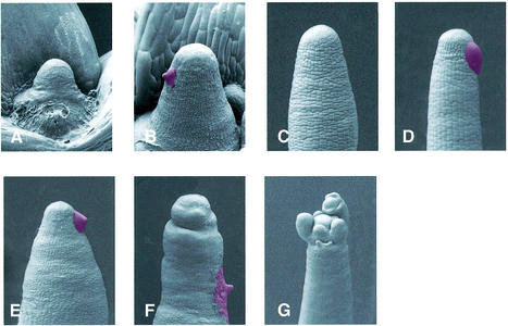
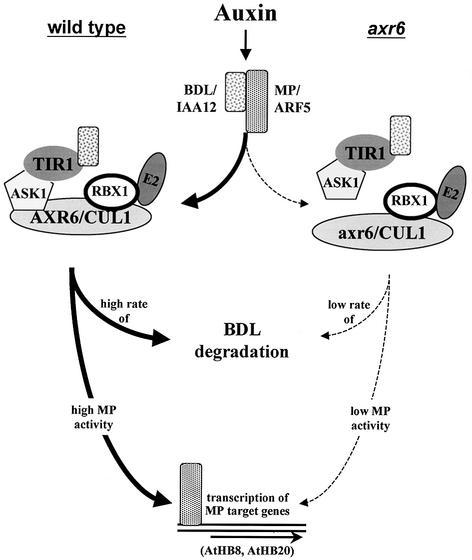
The AXR6 gene is required for auxin signaling in the Arabidopsis embryo and during postembryonic development. One of the effects of auxin is to stimulate degradation of the Aux/IAA auxin response proteins through the action of the ubiquitin protein ligase SCF(TIR1). Here we show that AXR6 encodes the SCF subunit CUL1. The axr6 mutations affect the ability of mutant CUL1 to assemble into stable SCF complexes resulting in reduced degradation of the SCF(TIR1) substrate AXR2/IAA7. In addition, we show that CUL1 is required for lateral organ initiation in the shoot apical meristem and the inflorescence meristem. These results indicate that the embryonic axr6 phenotype is related to a defect in SCF function and accumulation of Aux/IAA proteins such as BDL/IAA12. In addition, we show that CUL1 has a role in auxin response throughout the life cycle of the plant.


Figures

None
Fig. 1. Mapping of the AXR6 gene. (AAXR6 was mapped using crosses between axr6-2 (Col-0 background) and two mutants in the Ler background, ga1 and prl. The number of recombinants at each position for each cross are indicated. The mutation was localized to a region of ∼80 kb (indicated by the gray horizontal bar) on the short arm of chromosome 4 containing CUL1. (B) Sequencing of CUL1 in two independent alleles, axr6-1 and axr6-2, showed mutations in the same base at position 336. The mutations result in substitution of phenylalanine with valine and isoleucine in axr6-1 and axr6-2, respectively.
None
Fig. 2. CUL1 T-DNA insertion mutants are auxin resistant. (A) Two independent T-DNA insertion lines, cul1-3 and cul1-4 [Wasselewskja (WS) background], were identified carrying insertions in exon 15 of the gene. (B) Response of mutant and wild-type seedlings to auxin. At least 15 seedlings were tested for each genotype. Error bars represent standard deviation. (C) Heterozygous cul1-3/+ and cul1-4/+ plants have reduced CUL1 levels. Ten-day-old seedlings were used. The loading control (lower panel) shows an unidentified protein visualized by Poinceau staining.
None
Fig. 3. The axr6 mutants are deficient in auxin-induced gene expression and degradation of AXR2/IAA7. (A) RNA blot analysis shows that auxin-regulated expression of IAA5 and IAA7 is reduced in axr6 mutants. Seven-day-old seedlings were treated with buffer or 50 µM 2,4-D for 1 h before RNA extraction. CUL1 expression is not regulated by auxin and is not altered in axr6-1 or axr6-2 plants. Col-0 (lanes 1 and 4), axr6-1/+ (lanes 2 and 4), axr6-2/+ (lanes 3 and 6). (B) Pulse–chase analysis in 7-day-old seedlings showed that stability of the IAA7 protein is increased in axr6-1 and axr6-2. (C) The half-life of IAA7 is higher in the axr6 mutants. Values presented are the averages of three independent experiments ± SD. (P <0.05 for each mutant compared with wild type; Student’s t-test.)
None
Fig. 4. The axr6-1 and axr6-2 mutations alter CUL1 levels and formation of stable SCF complexes. (A) CUL1 levels in wild-type and mutant seedlings determined by western blot with antibody against CUL1. The lower panel shows an unidentified cross-reacting protein used as a loading control. (BIn vitro translation and GST–RUB1 modification of CUL1, AXR6-1 and AXR6-2 proteins in rabbit reticulocyte lysates. Arrow indicates GST–RUB1 modified cullin. (C) Effect of RBX1 over-expression on CUL1 levels in axr6 plants determined by western blot. (D) Co-immunoprecipitation of CUL1 (upper panel) and ASK1 (lower panel) from Col-0 and homozygous axr6-1 or axr6-2 seedlings. Proteins were immunoprecipitated with CUL1 antibody and analyzed by western blot. The ASK1 antiserum detected an unknown protein that was recovered after incubation with beads alone. (E) ASK1 levels in Col-0 and homozygous axr6-1 or axr6-2 extracts determined by western blot using α-ASK1 antibody. (F) Co-immunoprecipitation of CUL1 and RBX1 in Col-0 and mutant seedlings. (G) CUL1 levels in GST-IAA7 pulldowns from Col-0 and homozygous axr6 seedlings determined by western blot.
None
Fig. 5. Reduced CUL1 levels lead to severe developmental defects. Arabidopsis Col-0 plants were transformed with a CUL1 or CUL1K682M cDNA under the control of the 35S promoter from cauliflower mosaic virus. (A) Approximately 25% of the transgenic lines displayed growth defects of varying severity. The plant labeled ‘1’ is untransformed Col-0. The others are transgenic for 35S::CUL1K682M. (B) CUL1 levels in transgenic lines from (A) determined by western blot; 30 µg total protein were loaded in each lane. (C) Three-day-old kanamycin-resistant 35S::CUL1K682M seedlings with elongated hypocotyls were transferred to minimal medium lacking antibiotics and compared with Col-0 seedlings 12 days after transfer. Transgenic seedlings also displayed reduced root elongation and almost no lateral roots. (D) Surviving plants developed strongly distorted and compact inflorescences. (E) Compact inflorescence from a mature 35S::CUL1K682M plant. The frequency and severity of defects observed with the 35S::CUL1 construct were similar to those shown here.
None
Fig. 6. CUL1-deficient plants display defects in meristem growth and lateral organ initiation. (A) Stable CUL1-deficient plants exhibit slow and irregular leaf development. Leaf number was counted at intervals in Col-0 and two independent 35S::CUL1K682M lines. (B) Scanning electron micrograph of the SAM from a 35S::CUL1 seedling showing the pin phenotype. (C) Immunolocalization of CUL1 in Col-0 and 35S::CUL1K682M 9-day-old seedlings. Images from two different seedlings are shown for each genotype. The brown staining represents CUL1 protein. (D) Protein blot showing reduced CUL1 levels at the apex of 21-day-old plants. Thirty micrograms of total protein extract were loaded in each lane. (E) Pin-like meristem from inflorescences of 35S::CUL1K682 plants. (F) Homozygous axr1-13 carrying the 35S::CUL1K682 transgene generated by crossing a stable CUL1-deficient line(35S::CUL1K682) to axr1-13. All homozygous axr1-13 seedlings with at least one transgene had this phenotype. (G) Example of a T1 axr1-3 plant transformed with the 35S:CUL1 construct. Approximately 10% of the T1 seedlings had this phenotype.
None
Fig. 7. Response of CUL1-deficient pins to exogenous auxin. (A) Two-week-old 35S::CUL1K682 seedling with two cotyledons and two true leaves (one removed) and a central pin. (B) Arrested SAM 3 days after application of 1 mM IAA in lanolin paste (red). (C) Inflorescence pin on CUL1-deficient plant. (D) Inflorescence pin 3 days after local treatment with 1 mM IAA (red). (E) Inflorescences pin 3 days after local treatment with 1 mM IAA (red). (F) Floral pin 7 days after local treatment with 1 mM IAA (red). (G) Spontaneous flower formation on an inflorescence pin after prolonged growth.
None
Fig. 8. Role of AXR6/CUL1 during embryonic development. In wild-type plants (left side) embryonic development requires auxin-dependent degradation of BDL/IAA12 leading to transcription of MP target genes (Mattsson et al., 2003). MP transcriptional activators can act either as monomers or dimers (Tiwari et al., 2003). Possible target genes include AtHB8 and AtHB20 (Mattsson et al., 2003). In contrast, the axr6 mutations affect the interaction between ASK1 and CUL1 and reduce the number of functional SCF complexes (right side). As a result BDL–MP heterodimers are stabilized. Low levels of MP activity lead to abnormal embryonic development.

Similar articles

Cited by

References

    1. Baima S., Possenti,M., Matteucci,A., Wisman,E., Altamura,M.M., Ruberti,I. and Morelli,G. (2001) The Arabidopsis ATHB-8 HD-zip protein acts as a differentiation-promoting transcription factor of the vascular meristems. Plant Physiol., 126, 643–655. - PMC - PubMed
    1. Bennett S.R.M., Alvarez,J., Bossinger,G. and Smyth,D.R. (1995) Morphogenesis in pinoid mutants of Arabidopsis thaliana. Plant J., 8, 505–520.
    1. Berleth T. and Jurgens,G. (1993) The role of the monopteros gene in organising the basal body region of the Arabidopsis embryo. Development, 118, 575–587.
    1. Blilou I. et al. (2002) The Arabidopsis HOBBIT gene encodes a CDC27 homolog that links the plant cell cycle to progression of cell differentiation. Genes Dev., 16, 2566–2575. - PMC - PubMed
    1. Clough S.J. and Bent,A.F. (1998) Floral dip: a simplified method for Agrobacterium-mediated transformation of Arabidopsis thaliana. Plant J., 16, 735–743. - PubMed

Publication types