. 2003 Jul 1;22(13):3346-55.
doi: 10.1093/emboj/cdg322.

MEKK1 regulates calpain-dependent proteolysis of focal adhesion proteins for rear-end detachment of migrating fibroblasts

Affiliations

MEKK1 regulates calpain-dependent proteolysis of focal adhesion proteins for rear-end detachment of migrating fibroblasts

Bruce D Cuevas et al. EMBO J. .

Abstract

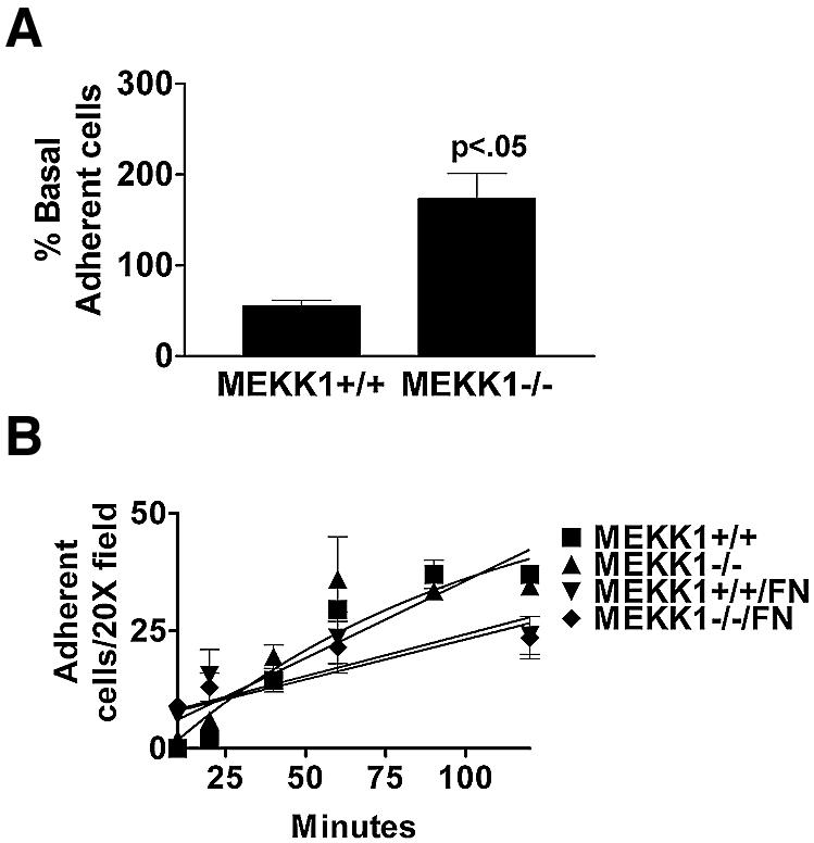
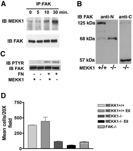
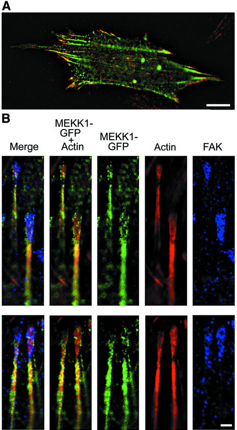
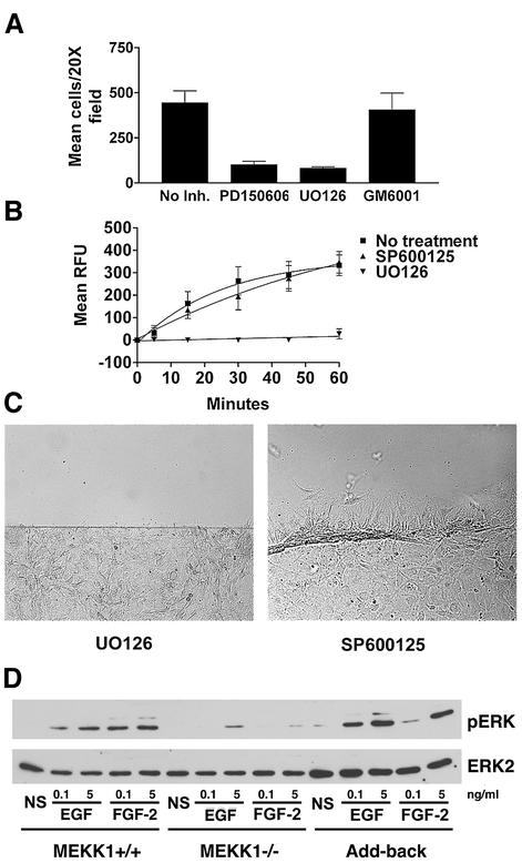
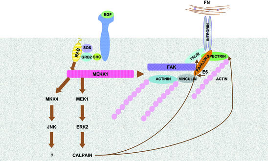
Herein, we define how MEKK1, a MAPK kinase kinase, regulates cell migration. MEKK1 is associated with actin fibers and focal adhesions, localizing MEKK1 to sites critical in the control of cell adhesion and migration. EGF-induced ERK1/2 activation and chemotaxis are inhibited in MEKK1-/- fibroblasts. MEKK1 deficiency causes loss of vinculin in focal adhesions of migrating cells, increased cell adhesion and impeded rear-end detachment. MEKK1 is required for activation of the cysteine protease calpain and cleavage of spectrin and talin, proteins linking focal adhesions to the cytoskeleton. Inhibition of ERK1/2 or calpain, but not of JNK, mimics MEKK1 deficiency. Therefore, MEKK1 regulates calpain-mediated substratum release of migrating fibroblasts.


Figures

None
Fig. 1. MEKK1-deficient fibroblasts show increased adherence characteristic of a defect in rear-end detachment. (A) Wild-type or MEKK1–/– MEFs were serum starved for 8 h, then treated with media with or without 10% fetal bovine serum (FBS). The plates were then inverted and centrifuged at 2300 g for 5 min. Adherent cells remaining attached to the well surface were stained with Wright’s stain and quantitated. Cell adherence is represented as the percent of the total serum-treated cells compared with the non-treated cells; 100% was taken as the number of wild-type cells in the dish before serum challenge, inversion and centrifugation. MEKK1–/– cells with serum challenge is >100% because more cells are retained after centrifugation than for the non-serum-stimulated wild-type cells, indicative of the increased adherence of MEKK1–/– cells. Results shown are the mean ± SEM of at least three independent experiments, and the statistical significance was determined by Student’s t-test. (B) Fibroblasts were resuspended in complete media and allowed to attach to either untreated or fibronectin-coated tissue culture plates. Cells were monitored for 2 h and the number of attached cells determined by phase microscopy. A digital movie of migrating MEKK1+/+ and MEKK1–/– fibroblasts is available as Supplementary data.
None
Fig. 2. MEKK1 expression is necessary for fibroblast migration. (A) Fibroblasts were seeded into the upper chamber of a Transwell migration plate with 5% FBS in the lower chamber. Cells traversed after 5 h to the lower surface of the membrane were quantitated. The results shown are the mean ± SEM of at least three independent experiments. (B) Fibroblasts were treated as in (A) except that the bottom well of the Transwell contained either 1 nM EGF, 100 µg/ml fibronectin or the combination of EGF and fibronectin. (C and E) Wild-type or MEKK1–/– fibroblasts were seeded onto coverslips and allowed to grow overnight. In addition, MEKK1–/– fibroblasts stably transfected with full-length MEKK1 (Add-back) were analyzed. Each confluent culture was ‘wounded’ with a razor and observed over the course of 5 h for migration into the wound space (in vitro wound healing assay). (C) is a DIC image of migrating cells. (D) The time required for confluent fibroblasts in a tissue culture plate to close a standardized wound (200 µm) is represented by the graph. Results shown are the mean ± SEM of at least three independent experiments, and the statistical significance was determined by Student’s t-test (*P < 0.05). (E) Cells were first stained with the fluorescent vital dyes PKH26 (red; MEKK1+/+) or PKH67 (green; MEKK1–/–) and then mixed in equal numbers before seeding onto coverslips, and treated as described above. The fluorescence image depicts the migration of MEKK1+/+ (wild type) and MEKK1–/– cells in co-culture. The data are representative samples from at least three independent experiments.
None
Fig. 3. Vinculin content in focal adhesions is diminished in migrating MEKK1–/– fibroblasts. A 0.4 µm deconvolved image section of the cell having the brightest focal adhesion staining was used for the measurement of integrated intensity of vinculin content for MEKK1+/+ (A), add-back (B) and MEKK1–/– (C) fibroblasts. The intensity of vinculin staining was measured per cell area of the section. The add-back clone stably expresses full-length MEKK1 and was derived from the MEKK1–/– fibroblasts. The bar graph in (D) shows the analysis from three experiments where 58 wild-type, 97 MEKK1–/– and 96 add-back cells were analyzed for integrated vinculin staining intensity per cell area. Vinculin content in the MEKK1–/– clone is diminished at a statistically significant level from wild-type MEKK1+/+ cells (*P < 0.0001) and add-back cells (**P < 0.001). Bar = 50 µm.
None
Fig. 4. MEKK1 and FAK form a complex in EGF-stimulated fibroblasts. (A) Wild-type mouse embryo fibroblasts were treated with 100 ng/ml EGF for the indicated times. After cell lysis, endogenous FAK was immunoprecipitated with anti-FAK antibody, and associated MEKK1 detected by MEKK1 immunoblotting. Total immunoprecipitated FAK was measured by anti-FAK immunoblotting using the same blots as that for MEKK1 analysis. (B) Wild-type (MEKK1+/+) and MEKK1–/– fibroblasts stably expressing papilloma virus E6/E7 proteins were immunoblotted for FAK protein expression using an antibody recognizing the N- or C-terminal domain of FAK. FAK is a 125 kDa protein with N- and C-terminal cleavage fragments of 68 and 57 kDa, respectively. (C) Serum-starved MEKK1+/+ or MEKK1–/– MEFs were allowed to adhere to fibronectin-coated bacterial plates for 30 min, and then lysed. Endogenous FAK was immunoprecipitated with anti-FAK antibodies, and the level of tyrosine-phosphorylated FAK determined by phosphotyrosine immunoblotting. The membrane was then stripped and total FAK was assessed by FAK immunoblotting. (D) MEKK1+/+ and MEKK1–/– fibroblasts ± E6/E7 protein expression were analyzed for migration in Transwell assays using 5% FBS as described for Figure 2A. *Inhibition of MEKK1–/– E6/E7 cell migration is statistically significant relative to FAK–/– cells (P < 0.05) and MEKK1–/– cells (P < 0.001).
None
Fig. 5. MEKK1 localizes to focal adhesions. MEKK1–/– fibroblasts were transfected with EGFP–MEKK1, incubated in serum-free media for 12 h, then processed as described in Materials and methods and subjected to immunofluorescence analysis. (A) An MEKK1–/– MEF transfected with EGFP–MEKK1 and treated with anti-FAK antibodies (FAK displayed as red fluorescence). Bar = 10 µm. (B) Displayed are two representative examples of co-localization of EGFP–MEKK1 with endogenous FAK (purple) and actin (red). Bar = 1 µm.
None
Fig. 6. MEKK1-deficient fibroblasts show reduced calpain activity, and calpain inhibition mimics MEKK1 deficiency. In vivo calpain activity was assessed in fibroblasts using the cell-permeable, fluorescent calpain substrate SLLVY-AMC (A and B) and by anti-spectrin or anti-talin immunoblotting (C). (A) MEKK1+/+ cells in the presence and absence of the calpain inhibitor PD150606 (50 µM), MEKK1–/– and MEKK1 add-back cells were used for measurement of calpain activity. (B) FAK+/+ and FAK–/– cells were used to measure calpain activity as in (A). (C) The anti-spectrin and anti-talin immunoblots were stripped and reprobed with anti-m-calpain antibodies to verify protein levels. The immunoblots are representative of at least three independent experiments. (D) Wild-type fibroblasts grown to confluency on coverslips were pre-treated for 1 h with 50 µM PD150606 (left panel) or 2 µM GM6001, a matrix metalloproteinase inhibitor (right panel), and then analyzed for migration using the in vitro wound healing assay following a razor swipe in the continuous presence of inhibitor. Results are representative of at least three independent experiments for each set of experiments in (A–D).
None
Fig. 7. Fibroblast migration and calpain activity are dependent on MEK, but not JNK activity. (A) Wild-type fibroblasts were loaded into Transwell migration chambers (105 cells/well) and allowed to migrate for 5 h, using 5% serum as a chemotactic agent. Calpain inhibitor PD150606 (50 µM), MEK inhibitor UO126 (10 µM) or matrix metalloproteinase inhibitor GM6001 (2 µM) were added to both the upper and lower chambers of the designated wells. (B) Wild-type fibroblasts were pre-incubated for 1 h with JNK inhibitor SP600125 (10 µM) or MEK inhibitor UO126 (10 µM), and calpain activity was assessed by SLLVY-AMC cleavage, and compared with that of non-treated cells. The results of both (A) and (B) are the mean ± SEM of at least three independent experiments. (C) Wild-type fibroblasts were pre-incubated with 10 µM SP600125 or 10 µM UO126 for 1 h and then analyzed for migration using the in vitro wound healing assay in the continuous presence of inhibitor. (D) Serum-starved wild-type, MEKK1–/– or MEKK1 add-back fibroblasts were treated with EGF or FGF-2 for 10 min and then lysed. ERK1/2 activation was then assessed by phospho-ERK immunoblotting. The membrane was then stripped and the total ERK2 level determined by ERK2 immunoblotting. The data are representative of at least three independent experiments. NS, no stimulus.
None
Fig. 8. Model depicting the FAK–MEKK1–MEK1/2–ERK2 pathway controlling calpain activation and the disruption of focal adhesion-actin cytoskeletal complexes. MEKK1–/– cells are defective in focal adhesion composition and regulation of the ERK2–calpain activation pathway (see text for details).

Similar articles

Cited by

References

    1. Arthur J.S., Elce,J.S., Hegadorn,C., Williams,K. and Greer,P.A. (2000) Disruption of the murine calpain small subunit gene, Capn4: calpain is essential for embryonic development but not for cell growth and division. Mol. Cell. Biol., 20, 4474–4481. - PMC - PubMed
    1. Beckerle M.C., Burridge,K., DeMartino,G.N. and Croall,D.E. (1987) Colocalization of calcium-dependent protease II and one of its substrates at sites of cell adhesion. Cell, 51, 569–577. - PubMed
    1. Bialkowska K., Kulkarni,S., Du,X., Goll,D.E., Saido,T.C. and Fox,J.E. (2000) Evidence that β3 integrin-induced Rac activation involves the calpain-dependent formation of integrin clusters that are distinct from the focal complexes and focal adhesions that form as Rac and RhoA become active. J. Cell Biol., 151, 685–696. - PMC - PubMed
    1. Cary L.A., Klinghoffer,R.A., Sachsenmaier,C. and Cooper,J.A. (2002) SRC catalytic but not scaffolding function is needed for integrin-regulated tyrosine phosphorylation, cell migration and cell spreading. Mol. Cell. Biol., 22, 2427–2440. - PMC - PubMed
    1. Christerson L.B., Vanderbilt,C.A. and Cobb,M.H. (1999) MEKK1 interacts with α-actinin and localizes to stress fibers and focal adhesions. Cell Motil. Cytoskeleton, 43, 186–198. - PubMed