. 2003 Jul 1;22(13):3421-30.
doi: 10.1093/emboj/cdg327.

An ADAR that edits transcripts encoding ion channel subunits functions as a dimer

Affiliations

An ADAR that edits transcripts encoding ion channel subunits functions as a dimer

Angela Gallo et al. EMBO J. .

Abstract

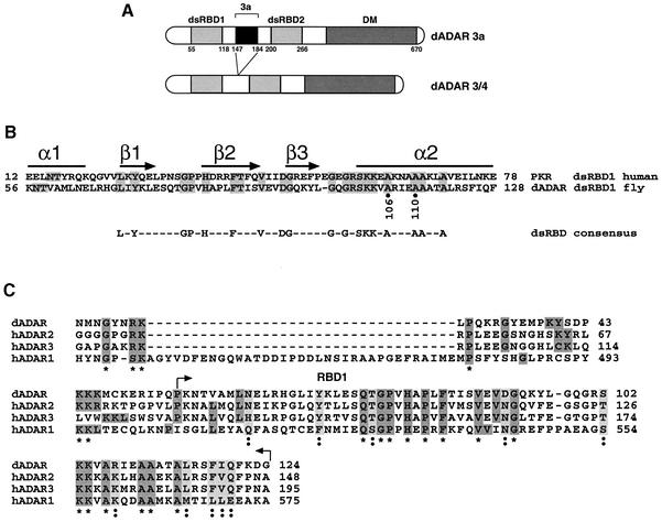
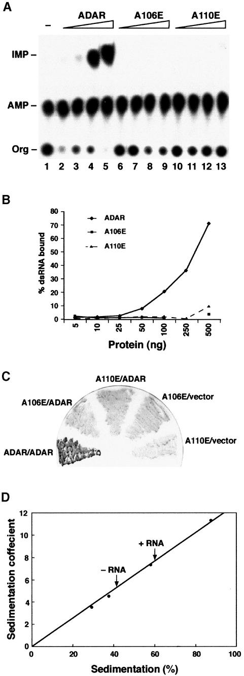
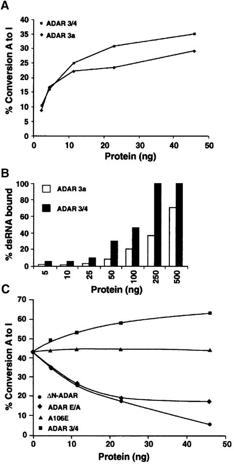
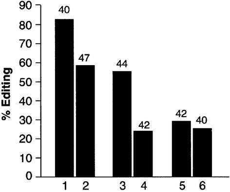
In this report, we establish that Drosophila ADAR (adenosine deaminase acting on RNA) forms a dimer on double-stranded (ds) RNA, a process essential for editing activity. The minimum region required for dimerization is the N-terminus and dsRNA-binding domain 1 (dsRBD1). Single point mutations within dsRBD1 abolish RNA-binding activity and dimer formation. These mutations and glycerol gradient analysis indicate that binding to dsRNA is important for dimerization. However, dimerization can be uncoupled from dsRNA-binding activity, as a deletion of the N-terminus (amino acids 1-46) yields a monomeric ADAR that retains the ability to bind dsRNA but is inactive in an editing assay, demonstrating that ADAR is only active as a dimer. Different isoforms of ADAR with different editing activities can form heterodimers and this can have a significant effect on editing in vitro as well as in vivo. We propose a model for ADAR dimerization whereby ADAR monomers first contact dsRNA; however, it is only when the second monomer binds and a dimer is formed that deamination occurs.


Figures

None
Fig. 1. Structure of Drosophila ADARs. (A) Schematic representation of the two dADAR isoforms, 3a and 3/4. The enzymes contain two dsRBDs and a deaminase domain (DM). The alternatively spliced exon 3a (111 nucleotides) lies between the two dsRBDs. Amino acid numbers are indicated below the domains. (B) Amino acid sequence comparison between dADAR and hPKR within the first dsRDB. The amino acids that are identical between the two proteins are in grey. A schematic representation of the predicted secondary structure of the dsRBD is shown above. The residues conserved >50% among all dsRBD sequences are shown beneath. (C) Homology within the minimum dimerization domain of the dADAR (AAF63703) to human ADARs (hADAR1-P55265, hADAR2-P78563, hADAR3-Q9NS39). This alignment was compiled with the web site http://ebiac.uk/clustalw. The alignment asterisks below represent identical amino acids and the double dots indicate similar amino acids.
None
Fig. 2. Dimerization of ADAR protein. (A) Yeast (L40) was cotransformed with plasmids encoding the full-length Adar fused to the LexA binding domain in the yeast expression vector pBTMK or fused to the DNA activation domain of GAL4 in the pACT2 vector. Positive protein–protein interactions were detected as blue colonies after a β-galactosidase assay. Full-length ADAR forms homodimers, and no interaction was detected with just the deaminase domain (DM) or with the controls. The positive control (C+) is the interaction between PAB1-2 and Paip 1, and the negative control (C-) is the absence of interaction between PAB1-2 and IRP (Gray et al., 2000). (B) The ADAR 3a isoform with epitope tags (c-Myc or HA) was transcribed and translated in vitro. The 3/4 isoform was generated with the anti-c-Myc epitope tag. The proteins were mixed and then co-immunoprecipitated with Myc monoclonal antibody. The immunoprecipitated proteins were electrophoresed on 8% SDS–polyacrylamide gel and revealed with an anti-HA polyclonal antibody. Recombinant ADAR 3a is able to form both homodimers and heterodimers.
None
Fig. 3. Minimum dimerization domain in ADAR protein. (A) Schematic representation of Adar deletions expressed in yeast as LexA fusion proteins. Terminal amino acid residue numbers are indicated. All the truncated proteins that retained the N-terminus expressed as LexA fused proteins interacted with full-length ADAR expressed as an AD fusion protein. A weak interaction was detected when the two dsRBDs were expressed as independent domains; blue colour appeared after 7–8 h incubation and is indicated as (–/+), strong interaction with blue colour after 20 min incubation is indicated as (++), and no interaction at any time incubation is indicated as (–). (B) Positive protein–protein interactions between ADAR and truncated ADAR proteins were detected as blue β-galactosidase activity by filter lift assay after 20 min. The amino acid residues encoding the truncated ADAR proteins are indicated. The black frame highlights the ΔN-ADAR mutant.
None
Fig. 4. dsRNA is required for ADAR dimerization. Single point mutations in dsRBD1 were generated so that amino acids A106 and A110 were changed to glutamate. (A) An in vitro editing assay was performed with recombinant proteins and chromatographed on a TLC plate. The positions of the origin (Org), adenosine (AMP) and inosine (IMP) are indicated. Increasing amounts of purified proteins were assayed [ADAR 3a (lanes 2–5), the mutant A106E (lanes 6–9) and the mutant A110E (lanes 10–13)], and the protein amounts were 10, 23, 70 and 140 ng, respectively. dsRNA without protein is in lane 1. (B) Filter binding was performed with the same mutant proteins A106E and A110E as well as with ADAR 3a; the protein amounts are indicated on the x-axis in nanograms. (C) Two-hybrid interactions between ADAR-LexA and ADAR-AD (positive control) and the two point mutants expressed as LexA fusion proteins and ADAR-AD were detected as blue β-galactosidase activity by filter lift assay. (D) Sedimentation coefficient of Drosophila ADAR 3/4-E/A in the presence or absence of RNA. Protein standards were applied to parallel gradients, and dADAR was detected by western blot analysis. The position of each protein is expressed as a percentage of the total number of fractions recovered from the gradient. The position of dADAR with and without RNA is indicated by arrows.
None
Fig. 5. ADAR is active as a dimer. (A) The in vitro editing assay was performed with either ADAR or ΔN-ADAR. Increasing amounts (7, 14, 30 and 50 ng) of both proteins were assayed. The products of the assay were chromatographed on a TLC plate and the origin (Org), adenosine (AMP) and inosine (IMP) are indicated. Lane 1 contains 32P-labelled dsRNA incubated without protein. Lanes 2–5 have increasing amounts of ADAR. Lanes 6–9 have increasing amounts of ΔN-ADAR. (B) Filter binding was performed with 32P-labelled dsRNA and increasing amounts of ADAR and ΔN-ADAR. The protein amounts are indicated on the x-axis in nanograms. This figure represents the average of two independent experiments.
None
Fig. 6. Inactive ADAR that retains the minimum dimerization domain downregulates ADAR activity in vitro. (A) A graph representing the editing activity of purified ADAR 3a and 3/4 on 32P-labelled dsRNA. The 3/4 isoform was more active than the 3a isoform. The protein amounts used are indicated. (B) A graph representing the binding of purified 3a and 3/4 proteins to 32P-labelled dsRNA. The 3/4 binding saturated at 250 ng, whereas the 3a isoform bound less dsRNA under the same experimental conditions. (CIn vitro editing assay was performed with a constant amount of ADAR 3a (7 ng). Increasing amounts of inactive 3/4-E/A (diamonds), or 3/4 wild-type (squares) or A106E (triangles) or ΔN-ADAR (circles) were added to this mixture; the protein amount that was added is indicated. A106E is a negative control as it cannot dimerize or bind to dsRNA, whereas ADAR 3/4 is a positive control.
None
Fig. 7. Formation of a heterodimer in Drosophila influences RNA- editing activity. Two independent RT–PCR reactions were performed on RNA isolated from transgenic flies containing both wild-type and the inactive ADAR 3/4-E/A. RNA editing at three different sites in two transcripts was analysed by sequencing individual clones. Lanes 1, 3 and 5 are sequences from wild-type Canton S, whereas lanes 2, 4 and 6 were obtained from the transgenic flies. Lanes 1 and 2 represent the percentage of editing at the S/G site in exon 15 in the cac transcript, lanes 3 and 4 the editing at the K/R site in the GluClα transcript, and lanes 5 and 6 the GluClα N/S site. The numbers above the bars in the graph are the numbers of clones sequenced.

Similar articles

Cited by

References

    1. Berg K.A., Cropper,J.D., Niswender,C.M., Sanders-Bush,E., Emeson,R.B. and Clarke,W.P. (2001) RNA-editing of the 5-HT2C receptor alters agonist-receptor-effector coupling specificity. Br. J. Pharmacol., 134, 386–392. - PMC - PubMed
    1. Betts L., Xiang,S., Short,S.A., Wolfenden,R. and Carter,C.W.Jr (1994) Cytidine deaminase. The 2.3 A crystal structure of an enzyme: transition-state analog complex. J. Mol. Biol., 235, 635–656. - PubMed
    1. Burns C.M., Chu,H., Rueter,S.M., Hutchinson,L.K., Canton,H., Sanders-Bush,E. and Emeson,R.B. (1997) Regulation of serotonin-2C receptor G-protein coupling by RNA editing. Nature, 387, 303–308. - PubMed
    1. Bycroft M., Grunert,S., Murzin,A.G., Proctor,M. and St Johnston,D. (1995) NMR solution structure of a dsRNA binding domain from Drosophila staufen protein reveals homology to the N-terminal domain of ribosomal protein S5. EMBO J., 14, 3563–3571. - PMC - PubMed
    1. Caceres J.F. and Kornblihtt,A.R. (2002) Alternative splicing: multiple control mechanisms and involvement in human disease. Trends Genet., 18, 186–193. - PubMed

Publication types