. 2003 Jul 1;22(13):3451-60.
doi: 10.1093/emboj/cdg320.

CRN-1, a Caenorhabditis elegans FEN-1 homologue, cooperates with CPS-6/EndoG to promote apoptotic DNA degradation

Affiliations

CRN-1, a Caenorhabditis elegans FEN-1 homologue, cooperates with CPS-6/EndoG to promote apoptotic DNA degradation

Jay Z Parrish et al. EMBO J. .

Abstract

Oligonucleosomal fragmentation of chromosomes in dying cells is a hallmark of apoptosis. Little is known about how it is executed or what cellular components are involved. We show that crn-1, a Caenorhabditis elegans homologue of human flap endonuclease-1 (FEN-1) that is normally involved in DNA replication and repair, is also important for apoptosis. Reduction of crn-1 activity by RNA interference resulted in cell death phenotypes similar to those displayed by a mutant lacking the mitochondrial endonuclease CPS-6/endonuclease G. CRN-1 localizes to nuclei and can associate and cooperate with CPS-6 to promote stepwise DNA fragmentation, utilizing the endonuclease activity of CPS-6 and both the 5'-3' exonuclease activity and a previously uncharacterized gap-dependent endonuclease activity of CRN-1. Our results suggest that CRN-1/FEN-1 may play a critical role in switching the state of cells from DNA replication/repair to DNA degradation during apoptosis.


Figures

None
Fig. 1. crn-1 encodes a FEN-1-like nuclear protein important for C.elegans apoptosis. (A) Alignment of CRN-1 and human FEN-1. Black shaded residues are identical and gray shaded residues are similar in two proteins. (B–E) TUNEL assays. N2 (B), cps-6(sm116) (C), nuc-1(e1392) (D) and ced-3(n2433) (E) animals were treated with control(RNAi) (filled bars) or crn-1(RNAi) (hatched bars) and their progeny were stained with TUNEL. The stages of embryos examined were comma, 1.5-fold and 3- and 4-fold. The y axis represents the mean number of TUNEL-positive cells present in the embryos (at least 12 embryos were scored at each stage). (F–I) Time course analysis of embryonic cell corpses. N2 (F), ced-8(n1891) (G), cps-6(sm116) (H), cps-6(sm116); ced-8(n1891) (I) animals were treated with control(RNAi) or crn-1(RNAi) and their progeny were scored for cell corpses in comma, 1.5-, 2-, 2.5-, 3-, 4-fold stage embryos and early L1 larvae. At least 15 animals were scored for each stage. (B to I) Data derived from control and crn-1(RNAi) treatment at the same stage were compared using unpaired t-test. *P < 0.05; **P < 0.002; ***P < 0.0001. All other points had P values > 0.05. Error bars indicate one standard deviation (SD). (J–M) Nuclear localization of CRN-1. Nomarski (J and L) and GFP fluorescent (K and M) images of a 1.5-fold stage transgenic embryo and a L1 transgenic larva are shown.
None
Fig. 2. Nuclease activities of CRN-1 and its interaction with CPS-6. (A) CRN-1 binds CPS-6. Purified GST or GST-fusion proteins (5 µg each) were used to precipitate [35S]methionine-labeled proteins as indicated. WT indicates the wild-type CPS-6 protein. ΔN denotes CPS-6(21–308). 30% of input [35S]methionine-labeled proteins is shown. (B) CRN-1 has flap endonuclease activity. FEN-1 or CRN-1 proteins (wild type or mutant) synthesized in the reticulocyte lysate were incubated with the labeled flap substrate, which is schematized below the image (lengths of oligonucleotides are indicated and * indicates the position of 32P-labeling). Cleavage products (19 and 21 nt) and their respective cleavage sites on the substrate (1 nucleotide 5′ or 3′ of the branch point) are indicated by arrows. WT, wild-type CRN-1; ED, CRN-1(E160D); DY, CRN-1(DY-AA). (C) CRN-1 possesses a previously uncharacterized gap-dependent endonuclease activity. A different labeled substrate was incubated with FEN-1 or CRN-1 proteins. The 19 nt endonucleolytic cleavage product and its corresponding cleavage site on the substrate are indicated with an arrow. The low-molecular-weight bands (indicated with arrowheads) observed at the bottom of the gel are products resulting from CRN-1 5′–3′ exonuclease digestion of the labeled 5′ blunt end. Neither CRN-1(E160D) nor CRN-1(DY-AA) is capable of generating these products since both lack the 5′–3′ exonuclease activity. (D) CRN-1 has 5′–3′ exonuclease activity. A 3′-end-labeled substrate was incubated with FEN-1 or CRN-1 proteins. The sizes of multiple cleavage products (indicated by arrows) are consistent with successive removal of 1 nt from the 5′ end of the labeled strand (indicated by an arrow) by the exonuclease. Reactions from panels B–D were resolved on 12% polyacrylamide/7 M urea gels and visualized using phosphorimager.
None
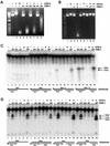
Fig. 3. CRN-1 and CPS-6 cooperate to promote DNA degradation. (A) Plasmid cleavage assay. CPS-6 (‘+’ denotes 50 ng, ‘++’ denotes 250 ng) or CRN-1 proteins (250 ng each) was incubated either alone or together as indicated with plasmid DNA (1 µg). WT, wild-type CRN-1; ED, CRN-1(E160D); DY, CRN-1(DY-AA). (B) Simultaneous presence of CRN-1 and CPS-6 is important for DNA degradation. In lanes 2–4, plasmid DNA was mock-treated with buffer and passed over Ni2+ NTA resin to simulate the depletion step. His6CPS-6 or CRN-1-His6 was subsequently incubated either alone or together with plasmid DNA for 30 min. In lanes 6–9, His6CPS-6 (lanes 6 and 8) or CRN-1-His6 (lanes 7 and 9) was first incubated with plasmid DNA for 30 min, depleted using Ni2+ NTA resin (1st), and plasmid DNA was subsequently incubated with CRN-1-His6 (2nd; lane 8) or His6CPS-6 (2nd; lane 9), or mock treated (buffer alone; ‘–’) for another 30 min. His6CPS-6 (50 ng) and 250 ng of CRN-1-His6 were used in all reactions. (C) CPS-6 enhances CRN-1 gap-dependent endonuclease activity. CRN-1 proteins (WT or mutants) or CPS-6 were incubated either alone or together, as indicated, with different substrates (schematized below reactions in which they were used). The endonucleolytic product sizes (indicated with arrows) increase with the increasing lengths of the single-stranded gaps in the substrates. (D) CPS-6 enhances CRN-1 5′–3′ exonuclease activity. Reactions were carried out as in (C) except that substrates were 3′-end labeled to monitor 5′–3′ exonuclease activity. Twelve, 11 and 10 nt exonucleolytic products were most prominent (indicated with arrows). In the presence of both CRN-1 and CPS-6, additional, smaller products were also visible. Reactions from panels C and D were resolved on 15% polyacrylamide/7 M urea gels and visualized using phosphorimager.
None
Fig. 4. Molecular model for CRN-1/CPS-6-mediated chromosome fragmentation during apoptosis. (A) Intact chromosomal DNA is likely nicked by CPS-6 (aided by CRN-1) and/or another nuclease (denoted by ?). (B) Following nicking, the 5′–3′ exonuclease activities of CRN-1 (aided by CPS-6), and possibly, other exonucleases (?) turn the nicks into gaps. (C) The resulting gapped substrates are cleaved by CRN-1 gap-dependent endonuclease activity (aided by CPS-6), resulting in fragmented substrates (D) which either are further processed by a 3′–5′ exonuclease(s) (?) (E) or can be directly processed (F) through similar steps (A–D) to generate smaller DNA fragments.

Similar articles

Cited by

References

    1. Bambara R.A., Murante,R.S. and Henricksen,L.A. (1997) Enzymes and reactions at the eukaryotic DNA replication fork. J. Biol. Chem., 272, 4647–4650. - PubMed
    1. Brenner S. (1974) The genetics of Caenorhabditis elegans. Genetics, 77, 71–94. - PMC - PubMed
    1. Enari M., Sakahira,H., Yokoyama,H., Okawa,K., Iwamatsu,A. and Nagata,S. (1998) A caspase-activated DNase that degrades DNA during apoptosis and its inhibitor ICAD. Nature, 391, 43–50. - PubMed
    1. Harrington J.J. and Lieber,M.R. (1994) The characterization of a mammalian DNA structure-specific endonuclease. EMBO J., 13, 1235–1246. - PMC - PubMed
    1. Horvitz H.R. (1999) Genetic control of programmed cell death in the nematode Caenorhabditis elegans. Cancer Res., 59, 1701s–1706s. - PubMed

Publication types

MeSH terms