

Review
doi: 10.1172/JCI19010.
Principles and practice of functional MRI of the human brain
Affiliations
- PMID: 12840051
- PMCID: PMC162295
- DOI: 10.1172/JCI19010
Item in Clipboard
Review
Principles and practice of functional MRI of the human brain
J Clin Invest.
2003 Jul.
No abstract available
Figures
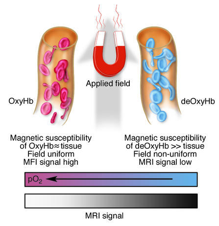
Schematic illustration of the origins of the BOLD effect in fMRI. While arterial blood is similar in its magnetic properties to tissue, deoxygenated blood is paramagnetic and so induces inhomogeneities within the magnetic field in tissue. These cause the MRI signal to decay faster. Signals from activated regions of cortex increase as the tissue becomes more magnetically uniform.
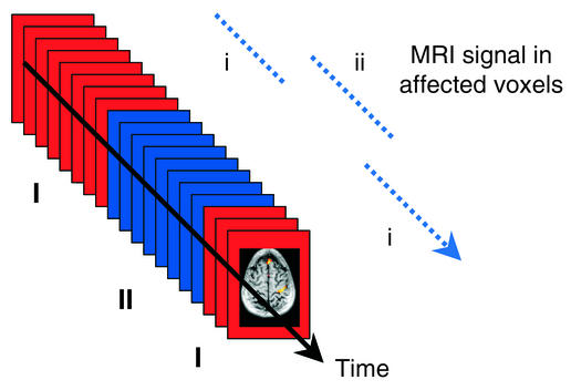
In a block design experiment, sequences of images are acquired in contrasting conditions (I and II). After acquisition, those voxels whose signals change in synchrony (i to ii) with the stimulus or task can be identified.
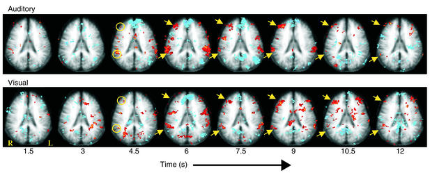
The average time course of the responses of one slice of the brain to oddball stimuli. Each image (left to right) represents the average time course of the response separated by 1.5 seconds, starting at the time of an auditory (top) or a visual (bottom) oddball stimulus. Various regions activate, and some of these activations are independent of the modality of the stimulus. The yellow arrows indicate activation in the middle frontal and supramarginal gyri. Adapted from ref. .
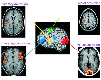
For surgical planning, a series of simple tasks (e.g., auditory, visual, motor, and language tasks) may be performed in a sequence to identify critical functional areas. These may then be superimposed on high-resolution anatomic images.
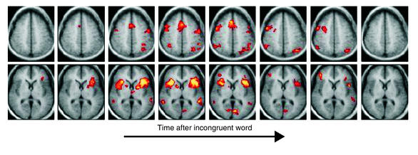
The average time course of the responses of two slices of the brain in the event-related Stroop task. Each image was acquired 1.65 seconds after the previous image, starting at the time of the incongruent word-color pair. Peak activities are apparent in the BOLD response 5–7 seconds after the event. A widespread pattern of activity is seen in both frontal and posterior regions, triggered by the Stroop effect. Adapted from ref. .
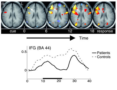
fMRI responses in prefrontal cortex during the rehearsal period of a verbal working memory task. Sustained BOLD effects are seen in normal subjects during the period 6–18 seconds after a series of words is encoded, but patients with schizophrenia fail to maintain this activity in inferior frontal gyrus (IFG), as shown in the lower time course. BA, Brodmann’s area.
References
-
- Robson M, Dorosz JL, Gore JC. Measurements of the temporal fMRI response of the human auditory cortex to trains of tones. Neuroimage. 1998;7:185–198. - PubMed
-
- Constable RT, McCarthy G, Allison T, Anderson AW, Gore JC. Functional brain imaging at 1.5T using conventional gradient echo MR imaging techniques. Magn. Reson. Imaging. 1993;11:451–459. - PubMed
-
- Cohen MS, Weisskoff RM. Ultra-fast imaging. Magn. Reson. Imaging. 1991;9:1–37. - PubMed
Publication types
MeSH terms
Substances
LinkOut - more resources
Full Text Sources
Other Literature Sources
Medical
