. 2003 Jul;112(1):67-75.
doi: 10.1172/JCI16645.

Role of host angiotensin II type 1 receptor in tumor angiogenesis and growth

Affiliations

Role of host angiotensin II type 1 receptor in tumor angiogenesis and growth

Kimiyasu Egami et al. J Clin Invest. 2003 Jul.

Abstract

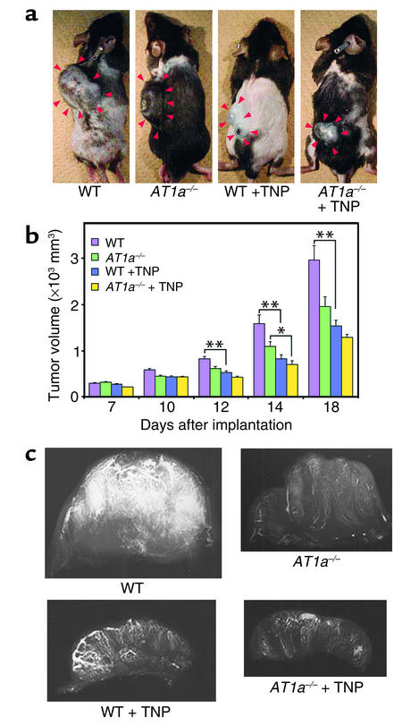
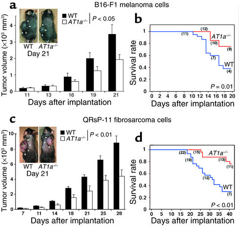
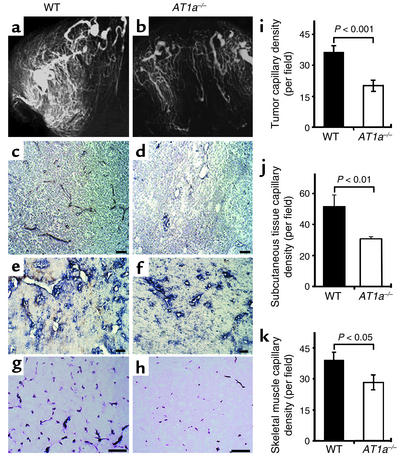
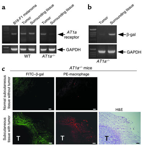
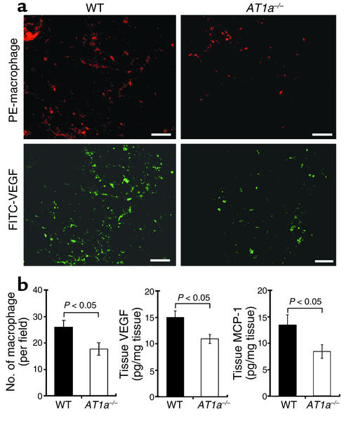
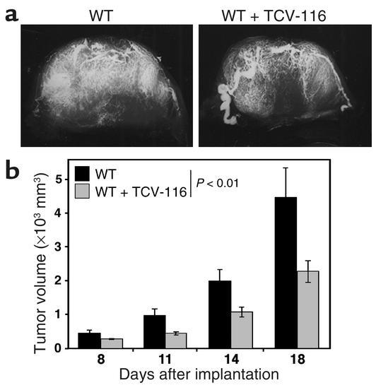
Although the renin angiotensin system (RAS) is a major regulator of vascular homeostasis, the role of the RAS in tumor angiogenesis is little understood. Here we show that host angiotensin II (ATII) type 1 (AT1) receptor plays an important role in angiogenesis and growth of tumor cells engrafted in mice. Subcutaneous B16-F1 melanoma-induced angiogenesis as assessed by tissue capillary density and microangiography was prominent in WT mice but was reduced in AT1a receptor-deficient (AT1a-/-) mice. Consequently, tumor growth rate was significantly slower, and the mouse survival rate was greater, in AT1a-/- mice than in WT mice. Tumor growth was also reduced in WT mice treated with TCV-116, a selective blocker of AT1 receptor. Because the beta-galactosidase gene was inserted into the AT1a gene locus in AT1a-/- mice, the site of beta-galactosidase expression represents the AT1a receptor expression in these mutant mice. In tumor-implanted AT1a-/- mice, the major site of the beta-galactosidase expression was macrophages in tissues surrounding tumors. Moreover, the number of infiltrated macrophages was significantly lower in AT1a-/- mice than in WT mice, and double-immunofluorescence staining revealed that these macrophages expressed VEGF protein intensively. Therefore, the host ATII-AT1 receptor pathway supports tumor-associated macrophage infiltration, which results in enhanced tissue VEGF protein levels. The host ATII-AT1 receptor pathway thereby plays important roles in tumor-related angiogenesis and growth in vivo.


Figures

Figure 1
Figure 1
Angiogenesis inhibitor TNP-470 suppresses tumor angiogenesis and growth. (a and b) A total of 106 B16-F1 melanoma cells were implanted subcutaneously into WT (n = 37) and AT1a–/– mice (n = 33) with or without TNP-470 administration. TNP-470 administration significantly inhibited tumor growth in both WT mice (n = 20) and AT1a–/– mice (n = 17). The inhibitory efficacy of TNP-470 was prominent in WT mice as compared with AT1a–/– mice. *P < 0.05; **P < 0.01. (c) Representative x-ray microangiograms of melanomas grown in WT and AT1a–/– mice with or without TNP-470. Administration of TNP-470 reduced angiographically visible tumor-related angiogenesis. TNP, TNP-470.
Figure 2
Figure 2
Host-derived AT1a receptor is important for tumor growth. (a) A total of 106 B16-F1 melanoma cells were implanted subcutaneously into WT (n = 11) and AT1a–/– (n = 12) mice. Tumor volumes were significantly smaller in the AT1a–/– group than in the WT group. (b) The Kaplan-Meier analysis shows the rate of survival was greater in the AT1a–/– group than in the WT group. Numbers in parentheses indicate the number of animals surviving at each time point. (c) A total of 4 × 10⁵ QRsP-11 fibrosarcoma cells were implanted subcutaneously into WT (n = 22) or AT1a–/– (n = 15) mice. Tumor volumes were significantly smaller in the AT1a–/– group than in the WT group. (d) The Kaplan-Meier analysis shows the rate of survival was greater in the AT1a–/– group than in the WT group. Numbers in parentheses indicate the number of animals surviving at each time point.
Figure 3
Figure 3
Decreased vascular density in and around B16-F1 melanomas implanted in AT1a–/– mice. Serial photo panels indicate representative x-ray microangiograms of melanomas grown in WT (a) or AT1a–/– (b) mice, CD31-stained melanoma mass grown in WT (c) or AT1a–/– mice (d) (×200), vWF-stained subcutaneous tissues surrounding B16-F1 melanomas grown in WT (e) or AT1a–/– mice (f) (×200), and alkaline phosphatase–stained skeletal muscle tissues underlying B16-F1 melanomas grown in WT (g) or AT1a–/– mice (h) (× 400). Bars indicate 50 μm. The capillary densities of (i) tumors, (j) subcutaneous tissue surrounding tumors, and (k) skeletal muscles underlying tumors were all significantly smaller in AT1a–/– mice (n = 6) than in WT mice (n = 6). Values represent the average number of vessels at ×200 ± the SE (by Student t test).
Figure 4
Figure 4
Host AT1a receptor is expressed on tumor-associated macrophages. (a) RT-PCR analysis for AT1a mRNA shows cultured B16-F1 melanoma cells, and implanted tumor tissues express AT1a mRNA. Subcutaneous tissues surrounding tumors expressed AT1a mRNA in WT mice but only slightly in AT1a–/– mice. (b) RT-PCR analysis for β-galactosidase (β-gal) mRNA in AT1a–/– mice shows subcutaneous tissues surrounding tumors express β-galactosidase (equivalent expression site of host AT1a receptor). β-Galactosidase mRNA is little expressed in tumors, indicating the absence of the host AT1a receptor within tumor tissues. (c) Subcutaneous tissues isolated from a remote normal skin and tumor-implanted site were stained with an FITC-conjugated anti–β-galactosidase mAb (representing host AT1a receptor) (FITC–β-gal) and phycoerythrin-conjugated anti-macrophage mAb (PE-macrophage). Panels indicate that macrophages located around tumors (TAMs) express β-galactosidase (host AT1a receptor). Bars indicate 100 μm. T, tumor.
Figure 5
Figure 5
TAMs express an angiogenic cytokine VEGF. (a) Macrophages were stained with a PE-conjugated anti-macrophage mAb (F4/80) in subcutaneous tissues surrounding tumors. Macrophages were costained with FITC-conjugated anti-VEGF mAb (FITC-VEGF). Bars indicate 50 μm. (b) Macrophages were counted under fluorescence microscopy (×200). The number of infiltrated macrophages was significantly lower in AT1a–/– mice (n = 5) than in WT mice (n = 5). Tissue VEGF and MCP-1 protein levels were significantly lower in AT1a–/– mice (n = 5) than in WT mice (n = 5).
Figure 6
Figure 6
Suppression of tumor angiogenesis and growth in WT mice by treatment with TCV-116, a selective AT1 receptor blocker. (a) Representative x-ray microangiograms of melanomas grown in WT mice with (right) or without (left) TCV-116. (b) A total of 106 B16-F1 melanoma cells were implanted subcutaneously into 33 WT mice with (n = 17) or without (n = 16) TCV-116 (10 mg/kg/day). Tumor volumes were calculated from tumor measurements scored at the indicated postimplantation day. The growth of B16-F1 melanoma was significantly reduced and delayed in WT mice treated with TCV-116 compared with control WT mice.

References

    1. Brunner HR, et al. Essential hypertension: rennin and aldosterone, heart attack and stroke. N. Engl. J. Med. 1972;286:441–449. - PubMed
    1. Lever AF, et al. Do inhibitors of angiotensin-I-converting enzyme protect against risk of cancer? Lancet. 1998;352:179–184. - PubMed
    1. Folkman J. Angiogenesis in cancer, vascular, rheumatoid and other disease. Nat. Med. 1995;1:27–31. - PubMed
    1. Hanahan D, Folkman J. Patterns and emerging mechanisms of the angiogenic switch during tumorigenesis. Cell. 1996;86:353–364. - PubMed
    1. Volpert OV, et al. Captopril inhibits angiogenesis and slows the growth of experimental tumors in rats. J. Clin. Invest. 1996;98:671–679. - PMC - PubMed

Publication types

MeSH terms