. 2003 Jul;112(1):83-90.
doi: 10.1172/JCI17845.

Transgenic amplification of glucocorticoid action in adipose tissue causes high blood pressure in mice

Affiliations

Transgenic amplification of glucocorticoid action in adipose tissue causes high blood pressure in mice

Hiroaki Masuzaki et al. J Clin Invest. 2003 Jul.

Abstract

Obesity is closely associated with the metabolic syndrome, a combination of disorders including insulin resistance, diabetes, dyslipidemia, and hypertension. A role for local glucocorticoid reamplification in obesity and the metabolic syndrome has been suggested. The enzyme 11beta-hydroxysteroid dehydrogenase type 1 (11beta-HSD1) regenerates active cortisol from inactive 11-keto forms, and aP2-HSD1 mice with relative transgenic overexpression of this enzyme in fat cells develop visceral obesity with insulin resistance and dyslipidemia. Here we report that aP2-HSD1 mice also have high arterial blood pressure (BP). The mice have increased sensitivity to dietary salt and increased plasma levels of angiotensinogen, angiotensin II, and aldosterone. This hypertension is abolished by selective angiotensin II receptor AT-1 antagonist at a low dose that does not affect BP in non-Tg littermates. These findings suggest that activation of the circulating renin-angiotensin system (RAS) develops in aP2-HSD1 mice. The long-term hypertension is further reflected by an appreciable hypertrophy and hyperplasia of the distal tubule epithelium of the nephron, resembling salt-sensitive or angiotensin II-mediated hypertension. Taken together, our findings suggest that overexpression of 11beta-HSD1 in fat is sufficient to cause salt-sensitive hypertension mediated by an activated RAS. The potential role of adipose 11beta-HSD1 in mediating critical features of the metabolic syndrome extends beyond obesity and metabolic complications to include the most central cardiovascular feature of this disorder.


Figures

Figure 1
Figure 1
(a) Daily profile of MAP in aP2-HSD1 mice at 23 weeks of age. MAP was monitored closely by telemetry transmitters implanted in the left carotid artery. Values are expressed as mean ± SEM of 60 data points each hour. Throughout the day, MAP in Tg mice (n = 17, filled squares) was significantly elevated (by 10–30 mmHg, P < 0.04) compared with that of non-Tg mice (n = 14, open circles). (b) In situ hybridization analysis of adrenal glands from aP2-HSD1 mice. Adrenal gland tissue from non-Tg (n = 5) and Tg mice (n = 3) was hybridized with 35S-labeled cRNA probes for aldosterone synthetase and 11β-hydroxylase. Images of five or six sections from each gland were quantified and statistically evaluated (d). Results are expressed as arbitrary units. The ratio of aldosterone synthetase to 11β-hydroxylase (Aldo/11β) in each sample was also determined. *P < 0.02 versus non-Tg. (c) Effect of specific AT-1 receptor antagonist GA0113 on MAP in aP2-HSD1 mice at 27 weeks of age. GA0113 was administered orally once per day (0.1 mg/kg body weight, at 1500 hours) for 4 days, and the effect of drug administration was evaluated by telemetry on day 5. Filled circles, non-Tg (n = 5) initial (untreated) values; open circles, non-Tg treated; filled squares, Tg (n = 6) initial (untreated); open squares, Tg treated. *P < 0.05 vs. Tg untreated. Telemetry monitoring was continued for 4 days after the final administration to observe that MAP in both sets of mice returned to initial values.
Figure 2
Figure 2
(a) Effect of high-salt diet on MAP in aP2-HSD1 mice. The aP2-HSD1 mice and non-Tg littermates at 19 weeks of age were given a high-salt (8%) diet for 3 weeks. The graph on the left shows that MAP in non-Tg mice (n = 4) fed a high-salt diet (filled circles) did not elevate significantly compared with the initial values when fed a chow diet with 1% salt (open circles). The graph at right shows that MAP in Tg mice fed a high-salt diet (filled squares) (n = 5) was significantly elevated during most of the day (except 0700 hours to 0800 hours, 1500 hours to 1600 hours, and 1800 hours to 2400 hours), by 10–20 mmHg compared with Tg mice fed a normal diet (open squares). *P < 0.05 vs. Tg mice fed a chow diet. (b) Effect of the specific AT-1 receptor antagonist GA0113 on MAP in aP2-HSD1 mice fed a high-salt diet. Four days of low-dose administration of GA0113 (0.1 mg/kg body weight, which does not lower MAP in non-Tg mice) to Tg mice fed a high-salt diet (22 weeks of age, n = 4) (open squares) markedly abrogated MAP elevation to approximately that of non-Tg mice (filled squares) fed chow diet. *P < 0.05 vs. Tg mice fed a high-salt diet.
Figure 3
Figure 3
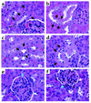
Histological examination of the kidneys of aP2-HSD1 mice and non-Tg littermates (24 weeks of age, male, n = 5 in each group). All are H&E stained sections photographed at an original magnification of ×400. (a and b) Distal tubular hyperplasia and hypertrophy with an origin immediately proximal to the macula densa are seen in Tg mice (b) compared with non-Tg littermates (a). The enlarged tubules in Tg mice (b, arrows) measured up to 2.5 times the cross-sectional area of tubules in non-Tg mice (a, arrows). (c and d) Distal tubular hypertrophy with increased cytoplasmic granularity and apical nuclear displacement are seen in Tg mice (d, arrows) compared with the normal, smaller distal tubules in non-Tg mice (c, arrows). (e and f) Moderate hyperplasia of the juxtaglomerular apparatus (JGA) at the vascular pole of the glomerulus is found in Tg mice (f) compared with non-Tg mice (e). White and blue arrows indicate glomeruli and JGAs, respectively. In both genotypes, glomeruli, proximal tubules, medullary thick ascending limb, and blood vessels were histologically unaltered.
Figure 4
Figure 4
Hypothetical representation of the pathophysiology of hypertension in aP2-HSD1 mice. Along with causing hyperinsulinemia and hyperleptinemia, reamplification of glucocorticoid action in adipose tissue leads to the activation of the circulating RAS and results in blood pressure elevation. This mouse model may provide a unique experimental system to better understand the pathophysiology of hypertension in human metabolic syndrome.

References

    1. Spiegelman BM, Flier JS. Obesity and the regulation of energy balance. Cell. 2001;104:531–543. - PubMed
    1. Matsuzawa Y, Funahashi T, Nakamura T. Molecular mechanism of metabolic syndrome X: contributions of adipocytokines, adipocyte-derived bioactive substances. Ann. N. Y. Acad. Sci. 1999;892:146–154. - PubMed
    1. Kahn BB, Flier JS. Obesity and insulin resistance. J. Clin. Invest. 2000;106:473–481. - PMC - PubMed
    1. Aizawa-Abe M, et al. Pathophysiological role of leptin in obesity-related hypertension. J. Clin. Invest. 2000;105:1243–1252. - PMC - PubMed
    1. Busetto L. Visceral obesity and the metabolic syndrome. Effect of weight loss. Nutr. Metab. Cardiovasc. Dis. 2001;11:195–204. - PubMed

Publication types

Substances