. 2003 Jul;112(1):126-35.
doi: 10.1172/JCI17669.

Expansion of human SCID-repopulating cells under hypoxic conditions

Affiliations

Expansion of human SCID-repopulating cells under hypoxic conditions

Guénahel H Danet et al. J Clin Invest. 2003 Jul.

Abstract

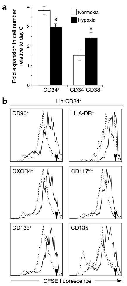
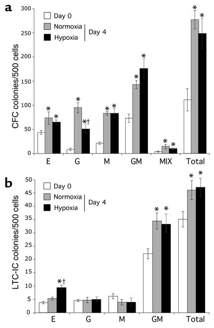
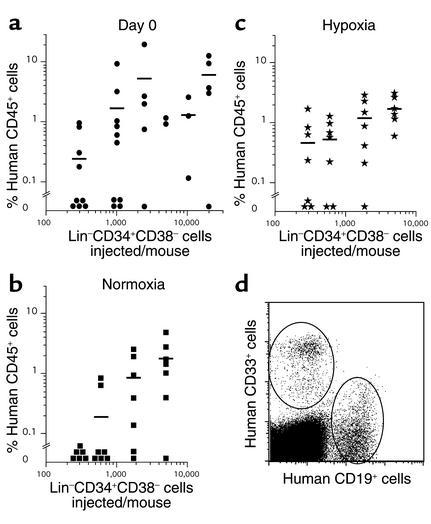
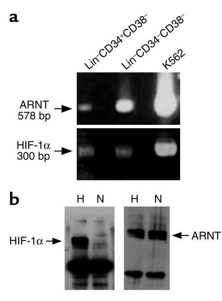
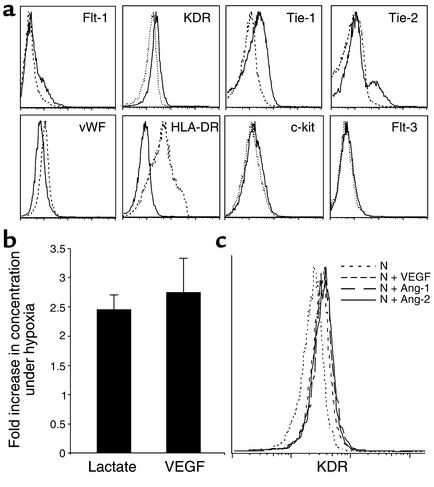
It has been proposed that bone marrow (BM) hematopoietic stem and progenitor cells are distributed along an oxygen (O2) gradient, where stem cells reside in the most hypoxic areas and proliferating progenitors are found in O2-rich areas. However, the effects of hypoxia on human hematopoietic stem cells (HSCs) have not been characterized. Our objective was to evaluate the functional and molecular responses of human BM progenitors and stem cells to hypoxic conditions. BM lineage-negative (Lin-) CD34+CD38- cells were cultured in serum-free medium under 1.5% O2 (hypoxia) or 20% O2 (normoxia) for 4 days. Using limiting dilution analysis, we demonstrate that the absolute number of SCID-repopulating cells (SRCs) increased by 5.8-fold in hypoxic cultures compared with normoxia, and by 4.2-fold compared with freshly isolated Lin-CD34+CD38- cells. The observed increase in BM-repopulating activity was associated with a preferential expansion of Lin-CD34+CD38- cells. We also demonstrate that, in response to hypoxia, hypoxia-inducible factor-1alpha protein was stabilized, surface expression of angiogenic receptors was upregulated, and VEGF secretion increased in BM Lin-CD34+ cultures. The use of low O2 levels to enhance the survival and/or self-renewal of human BM HSCs in vitro represents an important advance and could have valuable clinical implications.


Figures

Figure 1
Figure 1
Expansion and division history of BM cells after 4 days in culture in hypoxia or normoxia. LinCD34+ and LinCD34+CD38 cells were cultured in serum-free conditions for 4 days in the presence of IL-3, IL-6, SCF, Flt-3 ligand, and G-CSF. (a) Expansion. The fold increase in cell number relative to the initial cell number plated (day 0) is represented for each subpopulation cultured in normoxia (white bars) or hypoxia (black bars). *P < 0.05. (b) Division history of primitive subpopulations of LinCD34+ cells. Freshly isolated LinCD34+ cells were labeled with CFSE, cultured for 4 days in hypoxia (1.5% O2) or normoxia, and analyzed for CFSE fluorescence intensity and expression of markers associated with primitive progenitors and stem cells. Histograms (representative of three separate experiments) of CFSE fluorescence in various LinCD34+ cell subsets are shown after 4 days of culture in hypoxia (solid lines) or normoxia (dashed lines). Day 0 CFSE fluorescence intensity is indicated by an arrow on each histogram.
Figure 2
Figure 2
Effect of hypoxia on BM progenitor activity. The CFC (a) and LTC-IC (b) activity of BM LinCD34+CD38 cells was evaluated before (day 0; white bars) and after culture for 4 days under normoxic (gray bars) or hypoxic (1.5% O2; black bars) conditions. Data shown represent the number (mean ± SD) of BFU-E (E), granulocyte (G), monocyte (M), granulocyte-monocyte (GM), mixed (MIX, i.e., GM colonies with erythroid cells), and total number of colonies from three separate experiments. The paired Student t test was performed to compare day 0 with day 4 cells (*P < 0.05) and to compare hypoxic to normoxic conditions after 4 days of culture (P < 0.05).
Figure 3
Figure 3
Quantitative analysis of the effect of hypoxia on SRC frequency. The frequency of SRCs in freshly isolated LinCD34+CD38 cells (a, day 0) or cells cultured for 4 days under normoxic (b) or hypoxic (1.5% O2) (c) conditions was determined by limiting dilution analysis. For each NOD/SCID mouse (n = 88), the level of human engraftment (percentage of CD45+ cells) is represented as a function of the number of LinCD34+CD38 cells transplanted. (d) Representative flow cytometry dot plot showing the presence of both human myeloid (CD33+) and lymphoid (CD19+) cells in NOD/SCID BM 6–8 weeks after transplantation.
Figure 4
Figure 4
Expression of HIF in BM progenitors and stem cells. (a) RT-PCR analysis of ARNT and HIF-1α performed on RNA extracted from BM LinCD34+CD38, LinCD34CD38, and K562 cells (as a positive control). (b) Western blot analysis of cell extracts from BM LinCD34+ cells cultured overnight under hypoxic (H) or normoxic (N) conditions using antibodies against ARNT and HIF-1α. All gels are representative of at least three experiments.
Figure 5
Figure 5
Hypoxia regulates the expression of HIF target genes in BM progenitors and stem cells. (a) Representative histograms of cell surface expression of angiogenic and hematopoietic cytokine receptors in BM LinCD34+ cells after 4 days in culture under normoxic (dashed lines) or hypoxic (1.5% O2, solid lines) conditions. (b) The conditioned medium from BM LinCD34+ cells cultured overnight in normoxia or hypoxia (1.5% O2) was assayed for lactate and human VEGF. Bars represent the fold increase in lactate and VEGF concentrations measured in hypoxic cultures relative to normoxic conditions. Error bars show SD. (c) Effect of VEGF, angiopoietin-1 (Ang-1), and angiopoietin-2 (Ang-2) on cell surface expression of VEGFR2 in LinCD34+ cells after 4 days in culture under normoxic conditions (N).

References

    1. Harrison JS, Rameshwar P, Chang V, Bandari P. Oxygen saturation in the bone marrow of healthy volunteers. Blood. 2002;99:394. - PubMed
    1. Lichtman MA. The ultrastructure of the hemopoietic environment of the marrow: a review. Exp. Hematol. 1981;9:391–410. - PubMed
    1. Cipolleschi MG, Dello Sbarba P, Olivotto M. The role of hypoxia in the maintenance of hematopoietic stem cells. Blood. 1993;82:2031–2037. - PubMed
    1. Ivanovic Z, et al. Incubation of murine bone marrow cells in hypoxia ensures the maintenance of marrow-repopulating ability together with the expansion of committed progenitors. Br. J. Haematol. 2000;108:424–429. - PubMed
    1. Adelman DM, Maltepe E, Simon MC. Multilineage embryonic hematopoiesis requires hypoxic ARNT activity. Genes Dev. 1999;13:2478–2483. - PMC - PubMed