Case Reports
. 2003 Aug;73(2):355-69.
doi: 10.1086/377107. Epub 2003 Jul 1.

New molecular mechanism for Ullrich congenital muscular dystrophy: a heterozygous in-frame deletion in the COL6A1 gene causes a severe phenotype

Affiliations
Case Reports

New molecular mechanism for Ullrich congenital muscular dystrophy: a heterozygous in-frame deletion in the COL6A1 gene causes a severe phenotype

Te-Cheng Pan et al. Am J Hum Genet. 2003 Aug.

Abstract

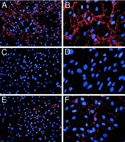
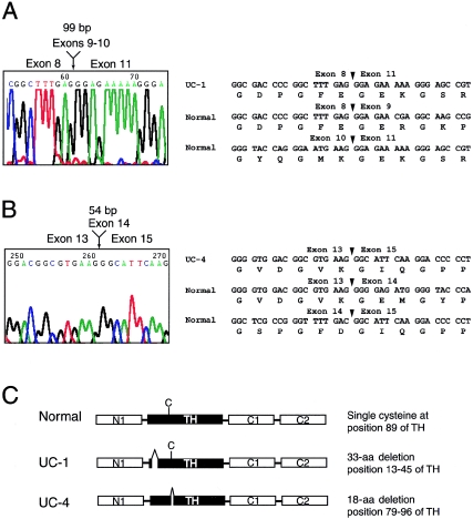
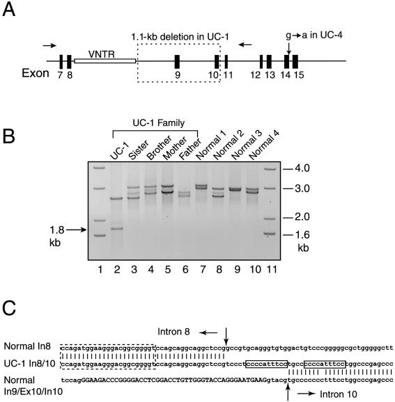
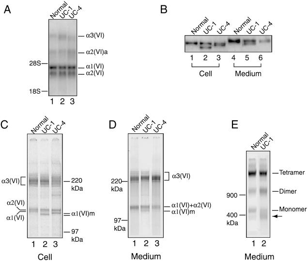
Recessive mutations in two of the three collagen VI genes, COL6A2 and COL6A3, have recently been shown to cause Ullrich congenital muscular dystrophy (UCMD), a frequently severe disorder characterized by congenital muscle weakness with joint contractures and coexisting distal joint hyperlaxity. Dominant mutations in all three collagen VI genes had previously been associated with the considerably milder Bethlem myopathy. Here we report that a de novo heterozygous deletion of the COL6A1 gene can also result in a severe phenotype of classical UCMD precluding ambulation. The internal gene deletion occurs near a minisatellite DNA sequence in intron 8 that removes 1.1 kb of genomic DNA encompassing exons 9 and 10. The resulting mutant chain contains a 33-amino acid deletion near the amino-terminus of the triple-helical domain but preserves a unique cysteine in the triple-helical domain important for dimer formation prior to secretion. Thus, dimer formation and secretion of abnormal tetramers can occur and exert a strong dominant negative effect on microfibrillar assembly, leading to a loss of normal localization of collagen VI in the basement membrane surrounding muscle fibers. Consistent with this mechanism was our analysis of a patient with a much milder phenotype, in whom we identified a previously described Bethlem myopathy heterozygous in-frame deletion of 18 amino acids somewhat downstream in the triple-helical domain, a result of exon 14 skipping in the COL6A1 gene. This deletion removes the crucial cysteine, so that dimer formation cannot occur and the abnormal molecule is not secreted, preventing the strong dominant negative effect. Our studies provide a biochemical insight into genotype-phenotype correlations in this group of disorders and establish that UCMD can be caused by dominantly acting mutations.


Figures

Figure  1
Figure 1
Deposition of collagen VI microfibrils in the extracellular matrix of dermal fibroblasts. Fibroblasts from an unaffected control individual (A and B), patient UC-1 (C and D), and patient UC-4 (E and F) were grown in the presence of 50 μg/ml L-ascorbic acid phosphate for 4 d postconfluency and were stained with the α1(VI) collagen-specific antibody. The antibody reaction was detected with Cy3-conjugated goat anti-rabbit IgG (red), and cells were counterstained with DAPI (blue) to visualize nuclei. Original magnifications ×20 (A, C, and E) and ×40 (B, D, and F).
Figure  2
Figure 2
In-frame deletions in the triple-helical domain of the COL6A1 mRNA in patients UC-1 and UC-4. A and B, DNA sequence chromatograms showing a deletion of 99 bp encoded by exons 9 and 10 in UC-1 (A) and a deletion of 54 bp encoded by exon 14 in UC-4 (B). DNA and amino acid sequences of the normal and mutant COL6A1 mRNA surrounding the mutations are shown. Arrowheads mark exon borders. C, Schematic diagram of normal and mutant α1(VI) collagen chains. The normal α1(VI) collagen chain consists of a triple-helical domain (TH) of 336 amino acids flanked by N- and C-terminal globular domains made up of vWF-A–like motifs (N1, C1, and C2). The TH domain contains a single cysteine residue (depicted as “C”) at position 89, which is important for dimer assembly. Patient UC-1 contains a 33–amino acid deletion near the N-terminus of the triple-helical domain, whereas patient UC-4 contains an 18–amino acid deletion somewhat downstream, which removes the cysteine residue.
Figure  3
Figure 3
Mutational analysis of the COL6A1 gene. A, Schematic diagram of the COL6A1 genomic region including exons 7–15 (blackened boxes). Exon 8 encodes the beginning of the triple-helical domain, and exons 9–15 each encode discrete numbers of Gly-Xaa-Yaa repeats in the triple-helical domain. A minisatellite (unblackened box labeled “VNTR”) is present at the 5′ end of intron 8. In patient UC-1, one of the COL6A1 alleles contains a 1.1-kb gene deletion (dotted box) extending from intron 8 to intron 10. Patient UC-4 carries a heterozygous G→A transition at the +1 position of intron 14. B, PCR amplification of genomic DNA from patient UC-1 and his family (lanes 2–6) and from four unaffected control individuals (lanes 7–10) with primers in introns 6 and 12 (arrows in A). Lanes 1 and 11 contain DNA size markers. C, DNA sequence of the 1.8-kb PCR product from UC-1, showing the breakpoint of the internal gene deletion (middle line) and its alignment with the normal COL6A1 genomic sequence of intron 8 (top line) and of intron 9–exon 10–intron 10 (bottom line). Exon sequences are in capital letters. Between the arrows are 15-bp inserted sequences in the deletion junction, which contains an 11-bp direct duplication of the sequence in intron 10 (boxed). The dotted box depicts the last repeat of the minisatellite sequence in intron 8. In = intron; Ex = exon.
Figure  4
Figure 4
Analysis of collagen VI mRNA and protein in fibroblasts from UC-1 and UC-4. A, Northern blot analysis of total RNA (5 μg) from normal fibroblasts and those from UC-1 and UC-4, with a mixture of [32P]dCTP-labeled cDNA probes for the three collagen VI mRNA. The probes hybridized with the α1(VI), α2(VI), and α3(VI) collagen mRNA of 4.2, 3.5, and 9–10 kb, respectively, plus an alternatively spliced α2(VI) collagen mRNA, α2(VI)a, of 6.0 kb. B, Immunoblot analysis of cell layer extracts (lanes 1–3) and culture media (lanes 4–6) from normal fibroblasts (lanes 1 and 4) and those from UC-1 (lanes 2 and 5) and UC-4 (lanes 3 and 6), with the antibody specific for the α1(VI) collagen chain. Lanes 1–3 contain 10 μg of total protein from the cell layers, and lanes 4–6 contain 100 μl of culture medium precipitated with 0.9 ml of 100% EtOH. C–E, Immunoprecipitation of cell layers (C) and culture media (D, E) from normal fibroblasts (lane 1) and those from UC-1 (lane 2) and UC-4 (lane 3), with the antibody specific for the α3(VI) collagen chain. Fibroblasts were labeled with [35S]cysteine overnight. Samples in B, C, and D were reduced with 25 mM DTT and separated on 3%–8% polyacrylamide gels. Samples in E were separated on a composite agarose polyacrylamide gel in the absence of 25 mM DTT. The mutant α1(VI) collagen chains are depicted as α1(VI)m. The arrow indicates a protein present in higher amounts in fibroblasts from UC-1 compared with the normal control fibroblasts.
Figure  5
Figure 5
Immunostaining of muscle biopsies from UC-1, UC-4, and an unaffected control individual. Sections were stained with the monoclonal antibody against collagen VI (green in A, D, and G) and the polyclonal antibody against perlecan (red in B, E, and H). C, F, and I show a merge of green and red fluorescence. In normal muscle (A, B, and C), collagen VI and perlecan colocalize in the basement membrane. In the muscle from UC-1 (D, E, and F), collagen VI is absent in the basement membrane surrounding muscle fibers; instead, it is accumulated in the interstitial and perivascular space between muscle fibers. In the muscle from UC-4 (G, H, and I), collagen VI is present in the basement membrane but appears patchy and significantly reduced relative to the perlecan staining. Collagen VI staining is increased in the interstitial and perivascular space.
Figure  6
Figure 6
Schematic diagram showing the effects of two different internal deletions on collagen VI assembly. A, Intracellular events. Normally (left), the α1(VI), α2(VI), and α3(VI) collagen chains presumably fold from the C-terminal end into a triple-helical monomer. Two key cysteine residues (vertical lines) in the N-terminal portion of the triple helix are critical for subsequent supramolecular assembly. Dimers are formed by the staggered antiparallel association of monomers with the N-terminal globular domains protruding at both ends. The outer cysteines contributed by either the α1(VI) or α2(VI) collagen chains are important for the dimer assembly. The dimers associate laterally into tetramers, which are stabilized by the outer cysteines contributed by the α3(VI) collagen chain. In UC-1 (middle) and UC-4 (right), equal amounts of normal and mutant (gray) α1(VI) collagen chains assemble into equal amounts of normal and mutant monomers. Because the cysteines critical for dimer and tetramer formation are preserved in UC-1, both normal and mutant monomers can assemble into dimers and tetramers in any combination. Thus, only 1/4 of the dimers and 1/16 of the tetramers are entirely composed of normal chains, whereas 15/16 of the tetramers contain a mixture of normal and mutant chains (gray striped lines). The deletion in UC-4 removes the cysteine in the triple-helical domain of the α1(VI) collagen chain, which prevents the abnormal monomers from assembling into dimers. Therefore, only normal tetramers are formed and secreted. The mutant chain in UC-4 may interfere with the assembly of the normal dimers, resulting in the formation and secretion of normal tetramers at a level ⩽1/2 that of the control. B, Extracellular event. Normally, tetramers are secreted and associated end-to-end into double-beaded microfibrils (top). The microfibrillar assembly involves interactions of the N- and C-globular domains and triple-helical domain. The deletion in UC-1 is located very close to the N-terminal end of the triple-helical domain (broken thick lines). Presumably, the triple helix in the mutant monomer is shortened because the normal α2(VI) and α3(VI) collagen chains are looped out at the region corresponding to the deletion (bottom). As a result, the mutant tetramers cannot properly align with normal tetramers to form the double-beaded structure. Because the majority of the secreted tetramers are abnormal, long microfibrils cannot be assembled.

References

Electronic-Database Information

    1. GenBank, http://www.ncbi.nlm.nih.gov/Genbank/ (for COL6A1 [accession numbers NM_0018489 and NT_011515], COL6A2 [accession numbers NM_001849 and NT_011515], and COL6A3 [accession numbers X52022 and NT_005120])
    1. Online Mendelian Inheritance in Man (OMIM), http://www.ncbi.nlm.nih.gov/Omim/ (for UCMD and Bethlem myopathy)

References

    1. Bachinger HP, Fessler LI, Timpl R, Fessler JH (1981) Chain assembly intermediate in the biosynthesis of type III procollagen in chick embryo blood vessels. J Biol Chem 256:13193–13199 - PubMed
    1. Bertini E, Pepe G (2002) Collagen type VI and related disorders: Bethlem myopathy and Ullrich scleroatonic muscular dystrophy. Eur J Paediatr Neurol 6:193–198 - PubMed
    1. Bethlem J, Wijngaarden GK (1976) Benign myopathy, with autosomal dominant inheritance: a report on three pedigrees. Brain 99:91–100 - PubMed
    1. Camacho Vanegas O, Bertini E, Zhang RZ, Petrini S, Minosse C, Sabatelli P, Giusti B, Chu ML, Pepe G (2001) Ullrich scleroatonic muscular dystrophy is caused by recessive mutations in collagen type VI. Proc Natl Acad Sci USA 98:7516–7521 - PMC - PubMed
    1. Camacho Vanegas O, Zhang RZ, Sabatelli P, Lattanzi G, Bencivenga P, Giusti B, Columbaro M, Chu ML, Merlini L, Pepe G (2002) Novel COL6A1 splicing mutation in a family affected by mild Bethlem myopathy. Muscle Nerve 25:513–519 - PubMed

Publication types

MeSH terms

Associated data