Comparative Study
. 2003 Jul 1;13(13):1106-15.
doi: 10.1016/s0960-9822(03)00418-4.

Rnd proteins function as RhoA antagonists by activating p190 RhoGAP

Affiliations
Comparative Study

Rnd proteins function as RhoA antagonists by activating p190 RhoGAP

Krister Wennerberg et al. Curr Biol. .

Abstract

Background: The Rnd proteins Rnd1, Rnd2, and Rnd3 (RhoE) comprise a unique branch of Rho-family G-proteins that lack intrinsic GTPase activity and consequently remain constitutively "active." Prior studies have suggested that Rnd proteins play pivotal roles in cell regulation by counteracting the biological functions of the RhoA GTPase, but the molecular basis for this antagonism is unknown. Possible mechanisms by which Rnd proteins could function as RhoA antagonists include sequestration of RhoA effector molecules, inhibition of guanine nucleotide exchange factors, and activation of GTPase-activating proteins (GAPs) for RhoA. However, effector molecules of Rnd proteins with such properties have not been identified.

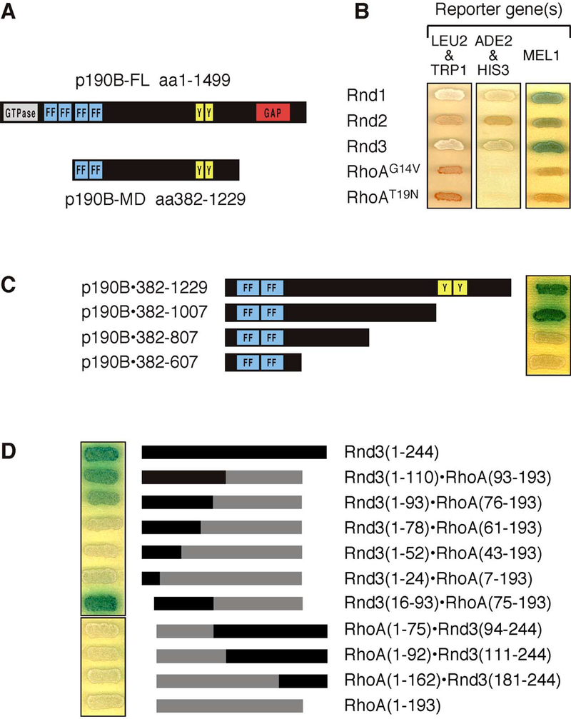
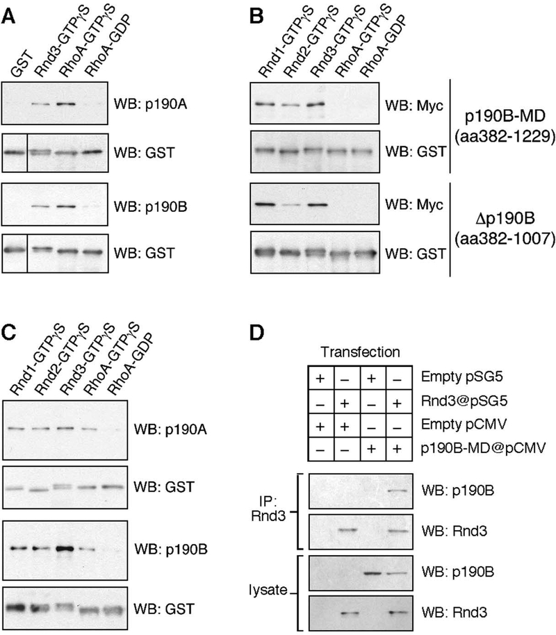
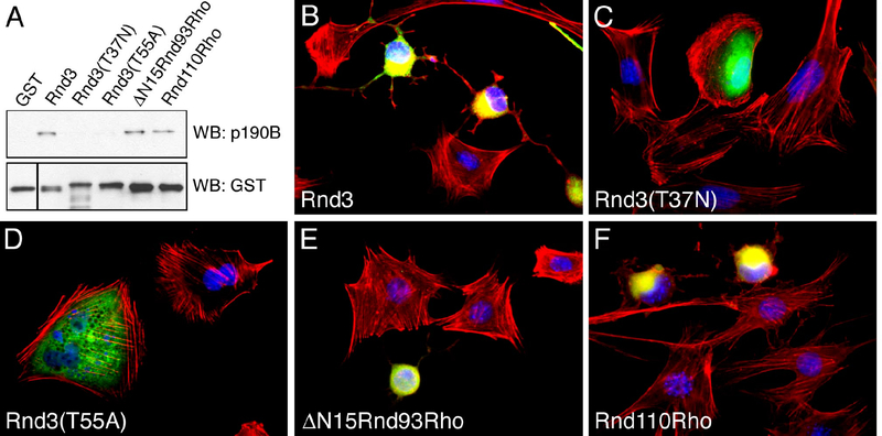
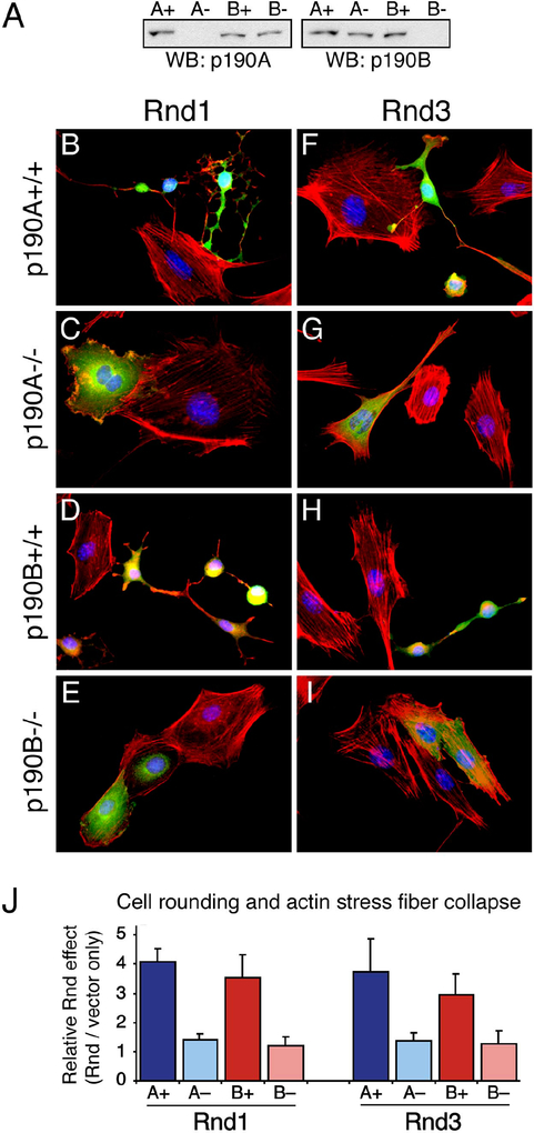
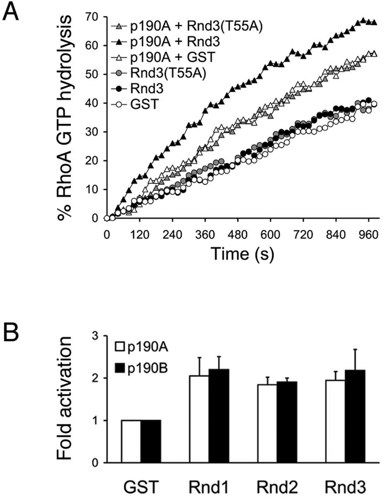
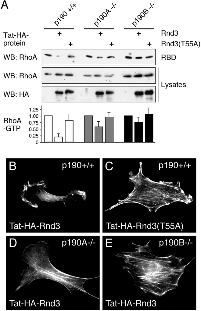
Results: Here we identify p190 RhoGAP (p190), the most abundant GAP for RhoA in cells, as an interactor with Rnd proteins and show that this interaction is mediated by a p190 region that is distinct from the GAP domain. Using Rnd3-RhoA chimeras and Rnd3 mutants defective in p190 binding, as well as p190-deficient cells, we demonstrate that the cellular effects of Rnd expression are mediated by p190. We moreover show that Rnd proteins increase the GAP activity of p190 toward GTP bound RhoA and, finally, demonstrate that expression of Rnd3 leads to reduced cellular levels of RhoA-GTP by a p190-dependent mechanism.

Conclusions: Our results identify p190 RhoGAPs as effectors of Rnd proteins and demonstrate a novel mechanism by which Rnd proteins function as antagonists of RhoA.


Figures

Figure 1:
Figure 1:. Rnd proteins interact with p190B-MD.
(A) Schematic presentation of full-length p190 and the fragment corresponding to p190B-MD isolated in the two-hybrid screen along with the known conserved domains found in these molecules, including the GTPase, FF, GAP, SH2 domain-binding (“Y”) domains. (B) Rnd1, Rnd2, and Rnd3, but neither constitutively active (G14V) nor dominant negative (T19N) RhoA, interact with p190B-MD in the two-hybrid assay as evidenced by activation of the HIS3 and ADE2 reporter genes (growth) as well as the MEL1 reporter (blue color). The orange-red color of the yeast grown on drop-out plates results from the AH109 yeast strain being auxotroph for adenine. (C) Truncation analysis of the Rnd3-interacting region of p190B-MD demonstrates that a fragment corresponding to amino acids 382–1007 is sufficient for mediating the interaction whereas further truncation of this fragment abrogates the activation of reporter genes. (D) Results obtained with RhoA-Rnd3 chimeras demonstrate that amino acids 16–93 in Rnd3 are required, and in the background of RhoA, sufficient to mediate the interaction with p190B-MD as demonstrated by activation of the MEL1 reporter gene. Note that for (B) and (C), Rnd proteins are expressed as fusion proteins with the Gal4 DNA-binding domain and the p190 derivatives as a fusion with the Gal4 activation domain, whereas in (D), the assay is reversed.
Figure 2:
Figure 2:. Interaction between Rnd and p190 molecules in GST pull-down and co-immunoprecipitation experiments.
(A) GST pull-down experiments with immobilized GST fusion proteins of Rnd3 loaded with GTPγS, or RhoA loaded with GTPγS or GDP, as well as GST alone demonstrates an interaction between Rnd3 and endogenous p190A or p190B. In the western blot to detect GST fusion proteins, the left lane has been shifted upwards to compensate for the higher mobility of non-fusion GST. (B) GST pull-down experiment from Cos7 cells transfected with expression constructs encoding Myc-epitope tagged p190B-MD (amino acids 382–1229), or a Myc-tagged fragment corresponding to amino acids 382–1007 of p190B. As shown, GTPγS-loaded Rnd1, Rnd2, and Rnd3, but not RhoA, bind p190B-MD or the 382–1007 fragment. (C) GST pull-down experiment with insect cell purified p190A and p190B demonstrating that the interaction between Rnd proteins and p190 molecules is direct. (D) Co-immunoprecipitation of p190B-MD with Rnd3 from lysates of Cos7 cells co-transfected with p190B-MD and Rnd3 expression vectors.
Figure 3:
Figure 3:. p190-binding correlates with phenotypic alterations elicited by Rnd3 derivatives.
(A) GST pull-down experiment from Cos7 cells transfected with p190B-MD expression construct. While GST-Rnd3, GST-ΔN15Rnd93Rho, and GST-Rnd110Rho all bind p190B-MD, no binding of GST-Rnd3(T37N), or GST-Rnd(T55A) to p190B-MD is observed. In the western blot to detect GST fusion proteins, the left lane has been shifted upwards to compensate for the higher mobility of non-fusion GST. (B–F) Effects on 3T3 fibroblasts associated with expression of Rnd3 and derivatives thereof as follows: (B) Rnd3; (C) Rnd3(T37N); (D) Rnd3(T55A); (E) ΔN15Rnd93Rho; (F) Rnd110Rho. Note that expression of ΔN15Rnd93Rho (E) or Rnd110Rho (F) elicits actin stress fiber collapse and cell rounding, effects associated with Rnd3 expression in 3T3 fibroblasts (B). In contrast, no alterations are observed following expression of Rnd3(T37N) (C), or Rnd3(T55A) (D). Cells were imaged for expression of Myc-tagged Rnd protein (green), filamentous actin (red), and nuclei (blue).
Figure 4:
Figure 4:. Attenuated phenotype of Rnd protein expression in cells deficient in p190A or p190B.
(A) Western blot analysis of p190 expression in whole cell lysates of 3T3 fibroblasts derived from p190A−/− or p190B−/− knock-out or littermate wild-type mice using antibodies specific for p190A (left panel) or p190B (right panel). (B–I) Phenotype elicited by expression of Rnd1 (B–E) or Rnd3 (F–I) in p190A control cells (B, F), p190A−/− cells (C, G), p190B control cells (C, H), or p190B−/− cells (E, I). The cells imaged for expression of Myc-tagged Rnd protein (green), filamentous actin (red), and nuclei (blue). While expression of Rnd1 or Rnd3 elicits cytoskeletal collapse and cell rounding in control 3T3 fibroblasts (B, D, F, H), the phenotype of Rnd1 or Rnd3 expression in p190A−/− as well as p190B−/− cells is substantially attenuated (C, E, G, I). (J) Quantification of actin stress fiber collapse and cell rounding in response to Rnd1 or Rnd3 expression in 3T3 fibroblasts deficient in p190A (A−) or p190B (B−), as well as in cells derived from littermate wild-type mice (A+; B+). For the quantification, cells transfected with Rnd expression vectors or control plasmid were identified by co-transfection with an EGFP expression vector to control for differences between the four cell lines in the endogenous level of actin stress fibers and cell rounding as detailed in Experimental Procedures. The data represent the mean and SEM of four independent experiments. Both microscopy and quantification were performed 36 hours after transfection.
Figure 5.
Figure 5.. Rnd proteins activate p190 molecules in vitro.
(A) In vitro GAP assays using fluorophore-labeled PBP to detect GTP hydrolysis. PBP and GTP-loaded RhoA was mixed with GST,GST-Rnd3, or GST-Rnd3(T55A) in the absence or presence of purified p190A (A), and the rates of RhoA GTP hydrolysis were measured as described in Experimental Procedures. (B) Activation of p190A- or p190B-mediated RhoA GTP hydrolysis by addition of GST-Rnd1, GST-Rnd2, or GST-Rnd3 (calculated as described in Experimental Procedures). The data shown represent the mean and SD of three independent experiments.
Figure 6.
Figure 6.. Rnd proteins activate p190 molecules in cells.
(A) Rhotekin RBD pulldown assay to determine levels of GTP-bound RhoA in control (p190A/B+/+), p190A−/−, and p190B−/− cells following transduction of Tat-HA-Rnd3 or Tat-HA-Rnd3(T55A). GTP-bound RhoA (RBD) and total RhoA in 2% of cell lysate (Lysates) were determined by western blot analysis using an antibody specific for RhoA. Protein transduction efficiency was confirmed with anti-HA antibody to detect Tat-HA-Rnd3 protein. The histogram shows RhoA-GTP levels in Tat-HA-Rnd3 and Tat-HA-Rnd3(T55A) transduced control, p190A−/−, and p190B−/− cells expressed as a ratio of RhoA-GTP levels in the corresponding untransduced cell lines. The data represent the mean and SD of three independent experiments. (B–E) Morphological effects of transduction of Tat-HA-Rnd3 (B, D, E) or Tat-HA-Rnd3(T55A) (C) into control cells (B and C) or cells deficient in p190A (D) or p190B (E).

References

    1. Takai Y, Sasaki T, Matozaki T (2001). Small GTP-binding proteins. Physiol. Rev 81, 153–208. - PubMed
    1. Foster R, Hu KQ, Lu Y, Nolan KM, Thissen J, Settleman J (1996). Identification of a novel human Rho protein with unusual properties: GTPase deficiency and in vivo farnesylation. Mol. Cell. Biol 16, 2689–2699. - PMC - PubMed
    1. Nobes CD, Lauritzen I, Mattei MG, Paris S, Hall A, Chardin P (1998). A new member of the Rho family, Rnd1, promotes disassembly of actin filament structures and loss of cell adhesion. J. Cell Biol 141, 187–197. - PMC - PubMed
    1. Hansen SH, Zegers MM, Woodrow M, Rodriguez-Viciana P, Chardin P, Mostov KE, McMahon M (2000). Induced expression of Rnd3 is associated with transformation of polarized epithelial cells by the Raf-MEK-extracellular signal- regulated kinase pathway. Mol. Cell. Biol 20, 9364–9375. - PMC - PubMed
    1. Guasch RM, Scambler P, Jones GE, Ridley AJ (1998). RhoE regulates actin cytoskeleton organization and cell migration. Mol. Cell. Biol 18, 4761–4771. - PMC - PubMed

Publication types

LinkOut - more resources