. 2003 Jul 1;17(13):1569-74.
doi: 10.1101/gad.1100303.

Werner syndrome protein limits MYC-induced cellular senescence

Affiliations

Werner syndrome protein limits MYC-induced cellular senescence

Carla Grandori et al. Genes Dev. .

Abstract

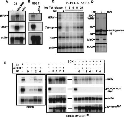
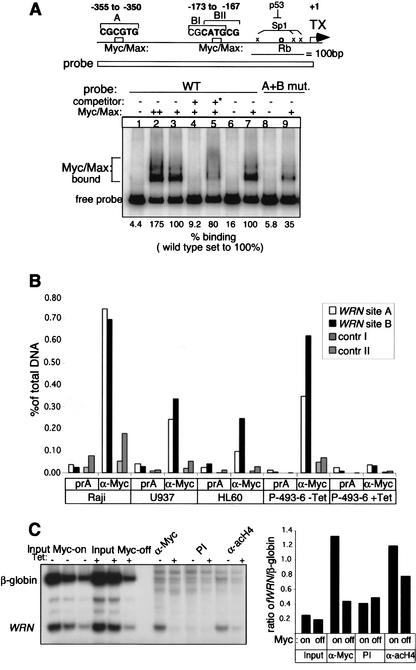
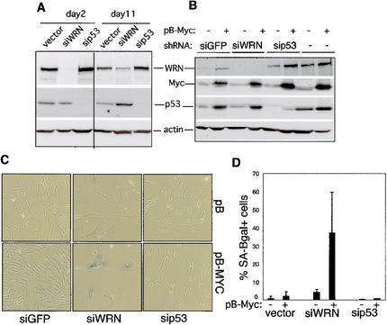
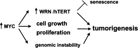
The MYC oncoprotein is a transcription factor that coordinates cell growth and division. MYC overexpression exacerbates genomic instability and sensitizes cells to apoptotic stimuli. Here we demonstrate that MYC directly stimulates transcription of the human Werner syndrome gene, WRN, which encodes a conserved RecQ helicase. Loss-of-function mutations in WRN lead to genomic instability, an elevated cancer risk, and premature cellular senescence. The overexpression of MYC in WRN syndrome fibroblasts or after WRN depletion from control fibroblasts led to rapid cellular senescence that could not be suppressed by hTERT expression. We propose that WRN up-regulation by MYC may promote MYC-driven tumorigenesis by preventing cellular senescence.


Figures

Figure 1.
Figure 1.
c-myc and WRN expression are tightly linked. (A) EBV-immortalized CB B cells overexpressing c-myc and max (+MycMax; Gu et al. 1993) have elevated WRN mRNAlevels compared with cells that overexpress max alone (+Max). (B) U937 leukemia cells show decreased levels of WRN and c-myc mRNAs after TPA-induced differentiation (U937+TPA). (C) Kinetics of WRN mRNAinduction following MYC up-regulation upon tetracycline withdrawal in B-cell line P-493-6 expressing Tet-myc (Schuhmacher et al. 1999). (D) Parallel induction of WRN and MYC protein in P-493-6 B cells labeled with 35S-methionine for 4 h after MYC induction: myc-on (–Tet), after growth for 48 h in the presence of tetracycline (myc-off; +Tet); and after 16 h of MYC induction: (16hr). Normalized lysates were immunoprecipitated with anti-WRN (Moser et al. 2000), anti-MAX (Santa Cruz Biotechnology no. sc765), or anti-MYC antibody (Santa Cruz Biotechnology no. sc764). (E) WRN induction by MYC does not require new protein synthesis. EREB B cells (Kempkes et al. 1995) expressing a chimeric MYC-ER™ protein (Wu et al. 1999) were grown in the absence of estrogen (E2–) to inactivate EBNA2-ER, then exposed for the indicated times to 4-OHT to induce MYC-ER™ in the presence or absence of cycloheximide. PhosphorImager quantitation of data in E indicated an approximately fivefold induction of WRN mRNA relative to actin by MYC-ER™ and approximately fourfold with cycloheximide, compared with an ∼1.2-fold increase due to cycloheximide addition to control EREB cells not expressing MYC-ER™.
Figure 2.
Figure 2.
In vitro and in vivo binding of MYC to the WRN promoter. (A) Purified MYC–MAX heterodimers bind and retard the mobility of a WRN promoter fragment encompassing nucleotides –412 to +8 in EMSAs. Where indicated, the A and B sites were mutated (see Materials and Methods) to determine the specificity of binding. In lane 2, 25 ng of MYC and 7.5 ng of MAX were used; In lanes 35, 7, and 9, 12.5 ng of MYC and 3.75 ng of MAX were used. Controls include a specific competitor oligonucleotide containing a MYC CACGTG consensus site added at 1 pM (∼10-fold molar excess over the probe; +), as well as a methylated CACGTG that is not recognized by MYC (*). (B) ChIP detects MYC binding to WRN promoter elements in vivo. Cross-linked chromatin from Raji, U937 and HL60 cell lines, and the EBV-immortalized P-493-6 cell expressing a Tet-myc was immunoprecipitated with protein Aonly (prA) or with anti-MYC antibodies. Bound DNAwas monitored by real-time PCR with WRN promoter-specific primers. Two negative controls were used as nontarget E-box-containing genes (see Materials and Methods). The percent of total DNAbound was calculated as described (Frank et al. 2001). (C) MYC induction increases MYC residence on the WRN promoter and correlates with increased histone H4 acetylation. ChIP assay using chromatin from P-493-6 cells expressing Tet-myc (–Tet; MYC-on), or from cells grown in the presence of tetracycline for 48 h (+Tet; MYC-off). Chromatin was immunoprecipitated with anti-MYC or anti-acetylated H4 (acH4; Upstate Biotechnology) antibodies or with rabbit preimmune serum. Bound DNAwas quantified by duplex PCR using primers flanking site B of the WRN promoter and the β-globin gene as internal control (Schubeler et al. 2000). The right panel shows the quantitation of the ratio of WRN promoter to β-globin. Similar results were obtained in three independent experiments.
Figure 3.
Figure 3.
Cells lacking WRN undergo rapid proliferative arrest and senescence upon c-myc overexpression. (A) hTERT-immortalized WRN–/– fibroblast strains AG00780 and AG03141 and control normal fibroblasts were transduced with LmycSN or LXSN (control) retrovirus; (Miller and Rosman 1989), then selected in G418. Approximately 30%–70% of WRN–/– infected with LmycSN showed morphological changes typical of senescent cells after 2–3 passages, and expressed SA-β-gal activity. (B) Proliferation of the cells as in A is indicated by the ratio of cells harvested/plated. (C) Western analysis of WRN and control cells at passage 3 indicates that c-myc overexpression leads to modest overexpression of p53 (Santa Cruz Biotechnology no. sc-126), p21Cip (BD Transduction Laboratories no. 610233), and p16ink4A (Pharmingen no. 13251A) in all cell types. WRN (BD Transduction Laboratories no. 611168) is undetectable in the WRN–/– lines as reported (Moser et al. 2000), but induced by MYC in control cells. (D) ARF mRNAexpression in WRN–/– and control hTERT+ fibroblasts was analyzed by Northern blot using an INK4a exon 1-specific probe. max was used as loading control. (E) hTERT mRNAexpression at passage 3 was quantitated by RT–PCR primers, and the ribosomal protein 36B4 was used as a control. HeLa and primary human foreskin fibroblasts (HFF) cDNAs were used as controls. The apparently higher levels of ARF mRNAin control cultures are due to RNAloading differences.
Figure 4.
Figure 4.
WRN depletion promotes MYC-induced senescence in normal hTERT+ fibroblasts. (A) Cells transduced with a control retroviral vector (LXSH) or with pBH-siWRN or pBH-sip53 were collected for Western analysis after 2 or 11 d of hygromycin selection (cells were passaged at day +7). Antibodies are as described in Figure 3B. Western analysis of simultaneous transduction of hTERT+ fibroblasts with pBH-siWRN and pB-MYC (+) or pBabe (–) retroviruses. Controls were pBH-siGFP or pBH-sip53. Cell lysates were collected after 3 d growth in the presence of hygromycin. (C) Representative fields of cells as in B, stained for SA-β-gal. (D) Percentage of SA-β-gal+ cells as in B after 7–9 d in hygromycin. Data are from three independent experiments, counting at least 5 different fields and/or 500 cells. As controls, the data obtained with pBH empty vector were pooled with pBH-siGFP.
Figure 5.
Figure 5.
Role of WRN induction in MYC-driven tumorigenesis. WRN appears to suppress genomic instability and cellular senescence by insuring the error-free repair of DNAfollowing damage or replication arrest. MYC overexpression promotes tumorigenesis by influencing cell growth, cell proliferation (for review, see Grandori et al. 2000), and genomic instability (Felsher and Bishop 1999; Vafa et al. 2002). MYC induction of WRN and hTERT (Wang et al. 1998a; Wu et al. 1999) may further promote tumorigenesis by suppressing cellular senescence while insuring a high probability of continued cell division.

References

    1. Adams J.M., Harris, A.W., Pinkert, C.A., Corcoran, L.M., Alexander, W.S., Cory, S., Palmiter, R.D., and Brinster, R.L. 1985. The c-myc oncogene driven by immunoglobulin enhancers induces lymphoid malignancy in transgenic mice. Nature 318: 533–538. - PubMed
    1. Alcorta D.A., Xiong, Y., Phelps, D., Hannon, G., Beach, D., and Barrett, J.C. 1996. Involvement of the cyclin-dependent kinase inhibitor p16 (INK4a) in replicative senescence of normal human fibroblasts. Proc. Natl. Acad. Sci. 93: 13742–13747. - PMC - PubMed
    1. Brosh R.M. and Bohr, V.A. 2002. Roles of the Werner syndrome protein in pathways required for maintenance of genome stability. Exp. Gerontol. 37: 491–506. - PubMed
    1. Brown J.P., Wei, W., and Sedivy, J.M. 1997. Bypass of senescence after disruption of p21CIP1/WAF1 gene in normal diploid human fibroblasts. Science 277: 831–834. - PubMed
    1. Brummelkamp T.R., Bernards, R., and Agami, R. 2002. A system for stable expression of short interfering RNAs in mammalian cells. Science 296: 550–553. - PubMed

Publication types

MeSH terms