. 2003 Jul 1;17(13):1646-62.
doi: 10.1101/gad.1100503.

Cell fate decisions within the mouse organizer are governed by graded Nodal signals

Affiliations

Cell fate decisions within the mouse organizer are governed by graded Nodal signals

Stephane D Vincent et al. Genes Dev. .

Abstract

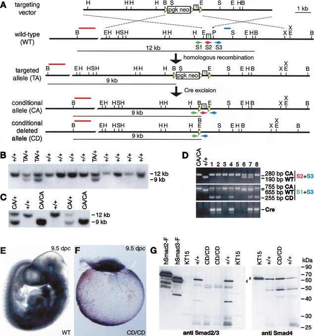
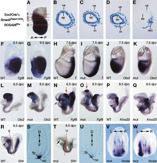
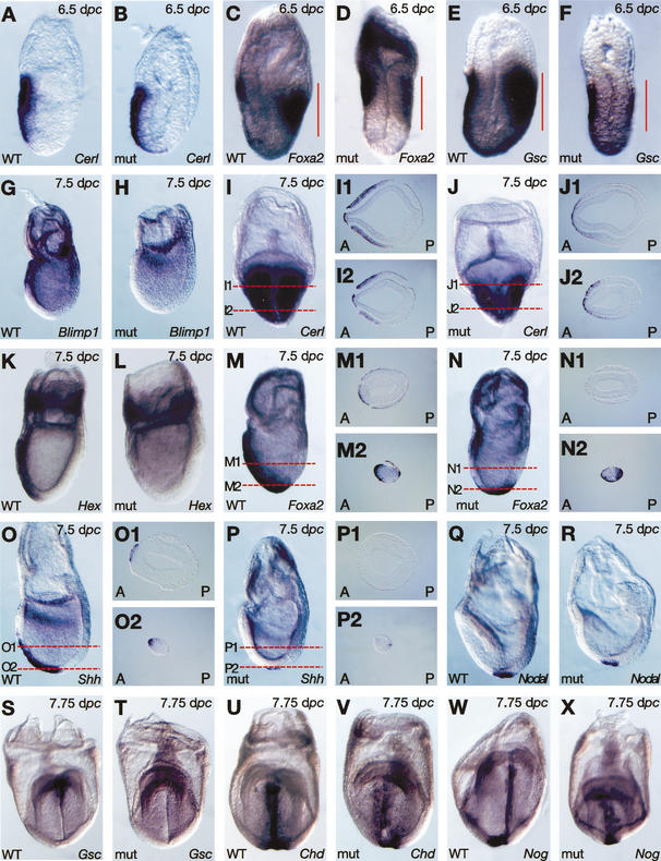
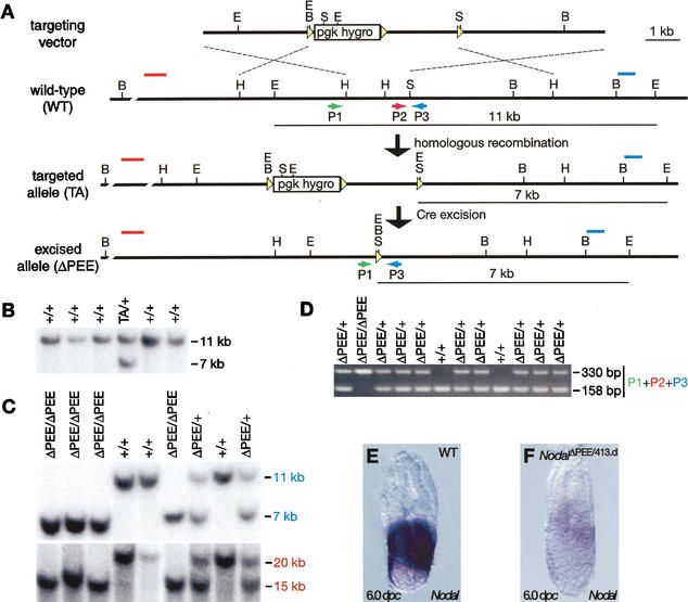
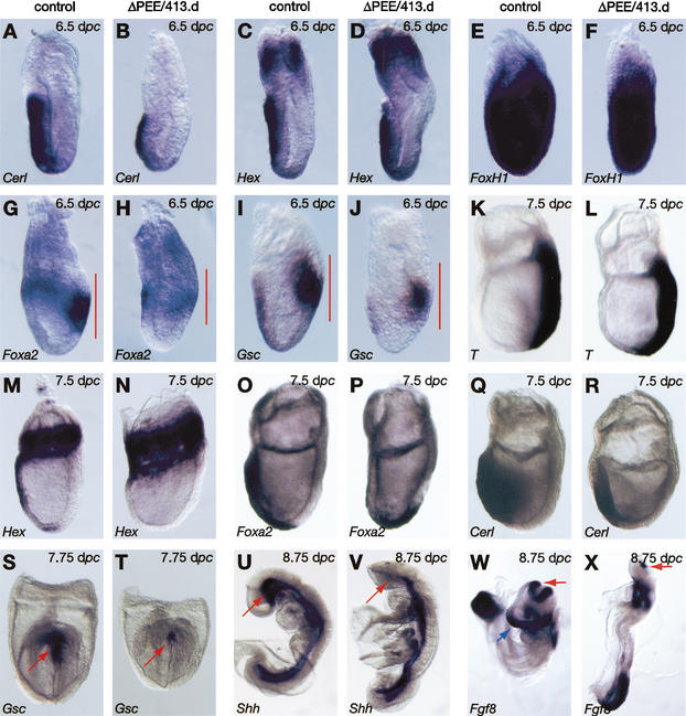
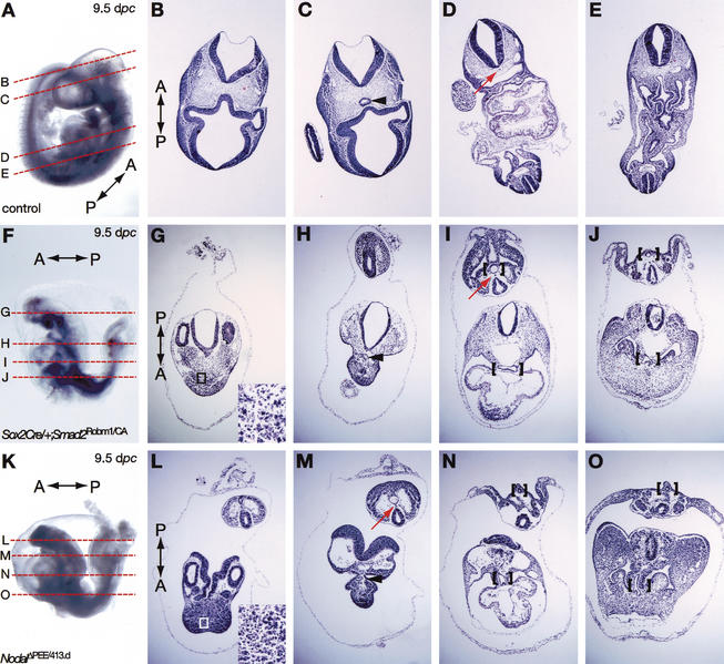
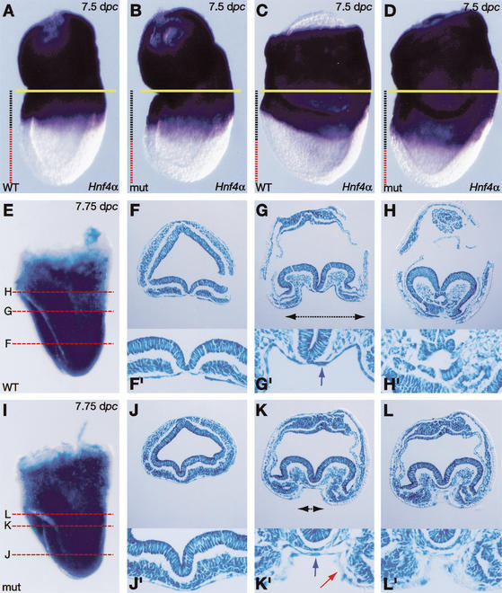
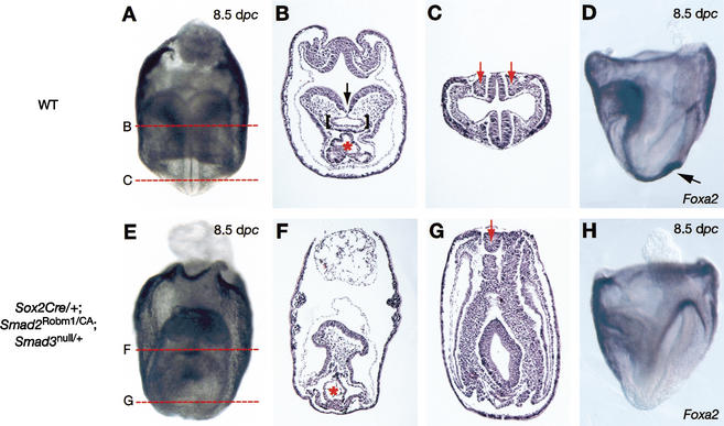
It is well known that cell fate decisions in the mouse organizer region during gastrulation ultimately govern gut formation and patterning, left-right axis determination, and development of the central nervous system. Previous studies suggest that signaling pathways activated by Nodal, bone morphogenetic protein (BMP), and Wnt ligands coordinately regulate patterning of the streak and the formation of midline organizing tissues, but the specific contributions of these molecules within discrete cell lineages are poorly defined. Here we removed Smad2 activity in the epiblast, using a conditional inactivation strategy. Abrogation of Smad2 does not compromise primitive streak (PS) formation or gastrulation movements, but rather results in a failure to correctly specify the anterior definitive endoderm (ADE) and prechordal plate (PCP) progenitors. To selectively lower Nodal activity in the posterior epiblast, we generated a novel allele lacking the proximal epiblast enhancer (PEE) governing Nodal expression in the PS. As for conditional inactivation of Smad2, germ-line deletion of the PEE selectively disrupts development of the anterior streak. In striking contrast, the node and its midline derivatives, the notochord and floor plate, develop normally in both categories of mutant embryos. Finally, we show that removal of one copy of Smad3 in the context of a Smad2-deficient epiblast results in a failure to specify all axial midline tissues. These findings conclusively demonstrate that graded Nodal/Smad2 signals govern allocation of the axial mesendoderm precursors that selectively give rise to the ADE and PCP mesoderm.


Figures

Figure 1.
Figure 1.
Generation and characterization of a Smad2 conditional allele. (A) Strategy used to flank the first coding exon of Smad2 with loxP sites. Targeted clones were identified using a 5′ external probe (red line) and an internal probe (blue line). Primers used for PCR are indicated by arrows. B, BamHI; E, EcoRI; H, HindIII; P, PstI (the asterisk indicates that a loxP site was introduced at this position); S, SpeI; X, XhoI. (B) Southern blot analysis of drug-resistant ES cell clones digested with BamHI and screened using the 5′ probe yields wild-type (WT; 12-kb) and targeted (9-kb) alleles. (C) Southern blot analysis of BamHI-digested tail DNAs from a Smad2CA/+ intercross litter. Smad2CA/CA animals are born at Mendelian ratios and are viable and fertile. (D) PCR analysis of tail-tip DNA samples from offspring from matings between Smad2CA/CA and Col2a1Cre/+ animals that express Cre in the cartilage (Ovchinnikov et al. 2000). Animals 1, 2, 4, 7, and 8carry the Cre transgene and the Smad2CD allele. (E) A 9.5-dpc wild-type (WT) embryo. (F) A 9.5-dpc Smad2CD/CD embryo composed only of extraembryonic tissues phenocopies Smad2Robm1/Robm1 embryos (Waldrip et al. 1998). (G) Western blot analysis with a monoclonal Smad2 antibody (left blot) and, as a control, a monoclonal Smad4 antibody (right blot). Smad2 and Smad3 are expressed by COS cells transfected with Smad2-Flag (hSmad2-F) or Smad3-Flag (hSmad3-F) expression constructs and 9.5-dpc wild-type (WT) yolk sacs, but no Smad2 signal was detected in Smad2Robm1/Robm1 KT15 ES cells or Smad2CD/CD embryo lysates. The positions of Smad2 (58kD) and Smad4 (66 kD) are indicated by arrowheads.
Figure 2.
Figure 2.
Deletion of Smad2 from the epiblast does not perturb gastrulation but leads to anterior patterning defects. (A) LacZ expression in 7.5-dpc Sox2Cre/+;Smad2CA/Robm1;ROSA26R/+ embryo. (BE) Sections of the embryo indicated in A. The extraembryonic ectoderm and visceral endoderm (VE) fail to express LacZ, whereas the epiblast and mesoderm are uniformly blue. ve, visceral endoderm; ep, epiblast; m, mesoderm; ee, extraembryonic ectoderm. The asterisk indicates the primitive streak. (FW) Whole-mount in situ hybridization of control (F,H,J,L,N,P,R) and Sox2Cre/+;Smad2CA/Robm1 mutants (G,I,K,M,O,Q,U). (FI) Streak formation and elongation proceed normally, as shown by Fgf8 and T expression. (J,K) Otx2, marker of the neural plate, is correctly expressed at 7.5 dpc. At early somite stages, the loss of anterior head structures is revealed by reduced expression of Otx2 (L,M) and by loss of the anterior neural ridge (blue arrow in N) corresponding to the most anterior Fgf8 domain (N,O). However, Fgf8 expression at the mid/hindbrain boundary (red arrow) is retained (N,O), and hindbrain formation is normal as assessed by Krox20 expression in rhombomeres 3 and 5 (P,Q). (R,T) Anterior truncation is also shown by the loss of the ventral domain of Shh (black arrow) in the brain. (S,U) In contrast, other Shh expression domains including the notochord (nc) and the floor plate (fp) are unaffected, as shown in frontal sections. (V,W) Nodal, normally confined to the left lateral plate mesoderm at the 3–5 somite stage, is bilaterally expressed in some mutant embryos. (AR,T) Lateral views with anterior to the left. (V,W) Ventral views. A–P, anterior–posterior axis; D–V, dorsal–ventral axis; L–R, left–right axis.
Figure 3.
Figure 3.
Mispatterning of the anterior streak derivatives in Sox2Cre/+;Smad2CA/Robm1 mutant embryos. Whole-mount in situ hybridization analysis of wild-type (WT) and Sox2Cre/+;Smad2CA/null mutant embryos at 6.5 dpc (AF), 7.5 dpc (GR), and 7.75 dpc (SX). (AD) As shown by Cerl and Foxa2 expression, the AVE is induced and rotates toward the presumptive anterior side of the embryo. (CF) However, the mutants express reduced levels of Foxa2 and Gsc in the anterior streak (indicated by red lines). Blimp1 normally expressed in the anterior mesendoderm (K) is absent in the mutant (L). Frontal views and transverse sections show that the Cerl expression domain in the definitive endoderm (DE) and mesoderm (I, sections I1 and I2) is reduced (J, sections J1, J2). Hex (K) and Foxa2 (M, section M1), normally expressed in the midline DE, are absent in the mutants (L,N, section N1). Note that Foxa2 is expressed in the node in both wild-type (WT; M, section M2) and mutant (N and section N2) embryos. As shown by Shh (O,P, sections O1 and P1, respectively) and Nodal (Q,R) expression domains, the node is formed normally. However, Shh (O, section O2) and Gsc (S) expression domains marking the prechordal plate (PCP) are missing in the mutant (P, cross-section P2; T). Chd and Nog normally expressed in the midline and PCP (U,W) are truncated anteriorly (V,X). A, anterior; P, posterior. Lateral views are shown with anterior to the left, with the exception of I, J, and SX, which show frontal views.
Figure 4.
Figure 4.
Generation of the NodalΔPEE allele. (A) Targeted deletion of the PEE element. Targeted clones identified using a 3′ external probe (blue line) were confirmed with a 5′ external probe (red line). The sizes of expected fragments are indicated. The arrows represent the primers used for genotyping. B, BamHI; E, EcoRI; H, HindIII; S, SpeI. (B) Southern blots of EcoRI-digested DNA from individual drug-resistant ES cell clones. The 3′ external probe detects 11-kb wild-type (WT) and 9-kb targeted alleles. (C) Southern blots of EcoRI- or BamHI-digested tail DNAs from intercross progeny using 3′ (blue) or 5′ (red) external probes, as indicated. (D) PCR genotyping screen of an intercross litter yields predicted 330-bp (mutant) and 158-bp (wild-type, WT) products. (E,F,G) Whole-mount in situ hybridization of wild-type (WT; E) and transheterozygous NodalΔPEE/413.d (F) 6.5-dpc embryos showing decreasing Nodal expression levels in the epiblast and VE.
Figure 5.
Figure 5.
NodalΔPEE/413.d mutants closely resemble Sox2Cre/+;Smad2CA/Robm1 mutant embryos. Whole-mount in situ analysis hybridization of wild-type (WT) and NodalΔPEE/413.d mutant embryos at 6.5 dpc (AJ), 7.5 dpc (KR), 7.75 dpc (S,T), 8.5 dpc (UV), and 8.75 dpc (WX). The AVE is specified and rotates toward the anterior side of the embryo (AD,GI), as shown by Cerl (A,B), Hex (C,D), Foxa2 (G,H), and Gsc (I,J) expression. Reduced Nodal activity has no effect on expression of FoxH1, a coeffector of Nodal signaling (E,F), or streak induction and elongation, as shown by T expression (K,L). As for the Smad2 conditional mutant embryos, expression of Foxa2 (G,H) and Gsc (I,J) is decreased in the anterior streak (indicated by red lines) compared with wild type (WT). Consequently, expression of the midline definitive endoderm markers Hex (M,N) and Foxa2 (O,P) is absent and the Cerl expression domain is reduced (Q,R). As for Smad2 conditional mutants, the NodalΔPEE/413.d mutants express Foxa2 in the forming node. (S,T) Frontal views show that Gsc is absent or severely reduced in the prechordal plate in NodalΔPEE/413.d mutant embryos. (U–W) Truncation of the anterior part of the brain is revealed at 8.5 dpc by the absence of the ventral forebrain domain of Shh (red arrow in U,V) and loss of Fgf8 marking the anterior neural ridge (blue arrow in W). (W,X) However, a residual Fgf8 isthmic region (red arrow) is still present in the mutants.
Figure 6.
Figure 6.
Gut tube formation in the NodalΔPEE/413.d and Sox2Cre/+;Smad2CA/Robm1 mutant embryos. Wild-type (WT; AE), Sox2Cre/+;Smad2CA/Robm1 mutant (FJ), and NodalΔPEE/413.d mutant (KO) 9.5-dpc embryos. (B,G,L) Sections at the level of the head show loss of anterior neurectoderm in both categories of mutant embryos and, as shown in high magnification in lower left (G,L), residual anterior tissue contains many pyknotic cells. (C) Normally the gut tube (arrowhead) extends into the head territories). In both classes of mutants (H,M) the anterior gut tube (arrowhead) is shortened and fails to extend anterior to the heart (D,I,N). Sections at the level of the heart reveal that the neural tube is highly abnormal. A poorly elaborated gut tube-like structure is present (brackets) but the heart fails to undergo correct looping morphogenesis and has an abnormal trifolium shape. (E,J,O) Sections at the level of the posterior somites. (E) In the wild-type (WT) embryo, the somites have already started to differentiate into dermomyotome and sclerotome. Distinct bilateral somites and hindgut (brackets) are present in both categories of mutant embryos. (I,M) The notochord (red arrow) is clearly detectable in the Smad2 and NodalΔPEE/413.d mutant embryos.
Figure 7.
Figure 7.
VE-derived cells contribute to the gut tube in Sox2Cre/+;Smad2CA/Robm1 mutants. (AD) Whole-mount in situ hybridization analysis of Hnf4α expression. In Smad2 conditional mutant embryos, the VE domain is incompletely displaced toward the proximal extraembryonic region. The boundary separating the extraembryonic and embryonic regions is indicated by a horizontal yellow line. The extent of Hnf4α expression relative to the distal nonstaining domain is indicated by black and red vertical lines, respectively. (EL) Sections of X-gal stained Sox2Cre/+;ROSA26R/+ and Sox2Cre/+;Smad2CA/Robm1;ROSA26R/+ (IL) 7.75-dpc embryos. The lateral limit of the definitive endoderm layer lining the foregut pocket (black arrows) is severely diminished in mutant (K) compared with wild-type (G) embryos. Wild-type definitive endoderm is exclusively composed of flattened LacZ-marked epiblast-derived cells (FH; magnification in F′–H′; blue arrow in G′), whereas the majority of cells in the mutant, especially in the most anterior part of the embryo, fail to express LacZ (K,L). (K′,L′) Higher-magnification views reveal the presence of intruding cuboidal endoderm (red arrow), morphologically similar to extraembryonic VE that predominates over midline epiblast-derived LacZ-marked cells (blue arrow in K).
Figure 8.
Figure 8.
Decreasing Smad3 expression in Smad2 conditional mutant embryos results in loss of axial structures. (A) Frontal view of a wild-type (WT) 8.5-dpc embryo showing the presence of a midline. (B,C) Hematoxylin-eosin (HE)-stained sections of A. The level of the sections is indicated in A. (E) Frontal view of a Sox2Cre/+;Smad2Robm1/CA;Smad3null/+ 8.5-dpc mutant embryo showing absence of midline structures. (FG) HE-stained sections of E. The level of sections is indicated in E. (D,H) Foxa2 whole-mount in situ analysis of wild-type (WT; D) and Sox2Cre/+;Smad2Robm1/CA;Smad3null/+ (H) 8.5 dpc embryos. (F) In the Sox2Cre/+;Smad2Robm1/CA;Smad3null/+ mutant embryo, the neural tissues remain as a neural plate, the heart (red asterisk) has not looped, and the foregut pocket (brackets in B) is absent. The absence of a midline is shown by the unfolded neural tube (arrow; cf. F and B) and the fused somites (red arrows; cf. G and C). Foxa2 is expressed in the node (arrow in D) and in the axial mesendoderm of the wild-type (WT) embryo. Neither of these expression domains is present in Sox2Cre/+;Smad2Robm1/CA;Smad3null/+ mutant embryos, confirming the absence of a node and its derivatives.

Similar articles

Cited by

References

    1. Agius E., Oelgeschlager, M., Wessely, O., Kemp, C., and De Robertis, E.M. 2000. Endodermal Nodal-related signals and mesoderm induction in Xenopus. Development 127: 1173–1183. - PMC - PubMed
    1. Ang S.L. and Rossant, J. 1994. HNF-3 β is essential for node and notochord formation in mouse development. Cell 78: 561–574. - PubMed
    1. Ang S.L., Conlon, R.A., Jin, O., and Rossant, J. 1994. Positive and negative signals from mesoderm regulate the expression of mouse Otx2 in ectoderm explants. Development 120: 2979–2989. - PubMed
    1. Bachiller D., Klingensmith, J., Kemp, C., Belo, J.A., Anderson, R.M., May, S.R., McMahon, J.A., McMahon, A.P., Harland, R.M., Rossant, J., et al. 2000. The organizer factors Chordin and Noggin are required for mouse forebrain development. Nature 403: 658–661. - PubMed
    1. Beddington R.S. and Robertson, E.J. 1999. Axis development and early asymmetry in mammals. Cell 96: 195–209. - PubMed

Publication types

MeSH terms