Detection and identification of ciprofloxacin-resistant Yersinia pestis by denaturing high-performance liquid chromatography
- PMID: 12843075
- PMCID: PMC165339
- DOI: 10.1128/JCM.41.7.3273-3283.2003
Detection and identification of ciprofloxacin-resistant Yersinia pestis by denaturing high-performance liquid chromatography
Abstract
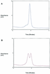
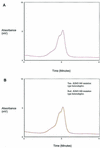
Denaturing high-performance liquid chromatography (DHPLC) has been used extensively to detect genetic variation. We used this method to detect and identify Yersinia pestis KIM5 ciprofloxacin-resistant isolates by analyzing the quinolone resistance-determining region (QRDR) of the gyrase A gene. Sequencing of the Y. pestis KIM5 strain gyrA QRDR from 55 ciprofloxacin-resistant isolates revealed five mutation types. We analyzed the gyrA QRDR by DHPLC to assess its ability to detect point mutations and to determine whether DHPLC peak profile analysis could be used as a molecular fingerprint. In addition to the five mutation types found in our ciprofloxacin-resistant isolates, several mutations in the QRDR were generated by site-directed mutagenesis and analyzed to further evaluate this method for the ability to detect QRDR mutations. Furthermore, a blind panel of 42 samples was analyzed by screening for two mutant types to evaluate the potential diagnostic value of this method. Our results showed that DHPLC is an efficient method for detecting mutations in genes that confer antibiotic resistance.
Figures


Similar articles
-
Detection of ciprofloxacin-resistant Yersinia pestis by fluorogenic PCR using the LightCycler.J Clin Microbiol. 2001 Oct;39(10):3649-55. doi: 10.1128/JCM.39.10.3649-3655.2001. J Clin Microbiol. 2001. PMID: 11574586 Free PMC article.
-
Identification of ciprofloxacin resistance by SimpleProbe, High Resolution Melt and Pyrosequencing nucleic acid analysis in biothreat agents: Bacillus anthracis, Yersinia pestis and Francisella tularensis.Mol Cell Probes. 2010 Jun;24(3):154-60. doi: 10.1016/j.mcp.2010.01.003. Epub 2010 Jan 25. Mol Cell Probes. 2010. PMID: 20100564
-
Detection of gyrA mutations in quinolone-resistant Salmonella enterica by denaturing high-performance liquid chromatography.J Clin Microbiol. 2002 Nov;40(11):4121-5. doi: 10.1128/JCM.40.11.4121-4125.2002. J Clin Microbiol. 2002. PMID: 12409384 Free PMC article.
-
Detection of mutations in gyrB using denaturing high performance liquid chromatography (DHPLC) among Salmonella enterica serovar Typhi and Paratyphi A.Trans R Soc Trop Med Hyg. 2016 Dec 1;110(12):684-689. doi: 10.1093/trstmh/trx002. Trans R Soc Trop Med Hyg. 2016. PMID: 28938049
-
Resistance of Yersinia pestis to antimicrobial agents.Antimicrob Agents Chemother. 2006 Oct;50(10):3233-6. doi: 10.1128/AAC.00306-06. Antimicrob Agents Chemother. 2006. PMID: 17005799 Free PMC article. Review. No abstract available.
Cited by
-
Effective plague vaccination via oral delivery of plant cells expressing F1-V antigens in chloroplasts.Infect Immun. 2008 Aug;76(8):3640-50. doi: 10.1128/IAI.00050-08. Epub 2008 May 27. Infect Immun. 2008. PMID: 18505806 Free PMC article.
-
Early indicators of exposure to biological threat agents using host gene profiles in peripheral blood mononuclear cells.BMC Infect Dis. 2008 Jul 30;8:104. doi: 10.1186/1471-2334-8-104. BMC Infect Dis. 2008. PMID: 18667072 Free PMC article.
-
A Survey of Antimicrobial Resistance Determinants in Category A Select Agents, Exempt Strains, and Near-Neighbor Species.Int J Mol Sci. 2020 Feb 29;21(5):1669. doi: 10.3390/ijms21051669. Int J Mol Sci. 2020. PMID: 32121349 Free PMC article.
-
Rapid genotyping of CTX-M extended-spectrum beta-lactamases by denaturing high-performance liquid chromatography.Antimicrob Agents Chemother. 2007 Apr;51(4):1446-54. doi: 10.1128/AAC.01088-06. Epub 2007 Jan 8. Antimicrob Agents Chemother. 2007. PMID: 17210774 Free PMC article.
-
Lovastatin protects against experimental plague in mice.PLoS One. 2010 Jun 2;5(6):e10928. doi: 10.1371/journal.pone.0010928. PLoS One. 2010. PMID: 20532198 Free PMC article.
References
-
- Arnold, N., E. Gross, U. Schwarz-Boeger, J. Pfisterer, W. Jonat, and M. Kiechle. 1999. A highly sensitive, fast, and economical technique for mutation analysis in hereditary breast and ovarian cancers. Hum. Mutat. 14:333-339. - PubMed
-
- Bauer, A. W., W. M. Kirby, J. C. Sherris, and M. Turck. 1966. Antibiotic susceptibility testing by a standardized single disk method. Am. J. Clin. Pathol. 45:493-496. - PubMed
-
- Cargill, M., D. Altshuler, J. Ireland, P. Sklar, K. Ardlie, N. Patil, N. Shaw, C. R. Lane, E. P. Lim, N. Kalyanaraman, J. Nemesh, L. Ziaugra, L. Friedland, A. Rolfe, J. Warrington, R. Lipshutz, G. Q. Daley, and E. S. Lander. 1999. Characterization of single-nucleotide polymorphisms in coding regions of human genes. Nat. Genet. 22:231-238. - PubMed
-
- Devaney, J. M., E. L. Pettit, S. G. Kaler, P. M. Vallone, J. M. Butler, and M. A. Marino. 2001. Genotyping of two mutations in the HFE gene using single-base extension and high-performance liquid chromatography. Anal. Chem. 73:620-624. - PubMed
Publication types
MeSH terms
Substances
Associated data
- Actions
LinkOut - more resources
Full Text Sources

