. 2003 Sep 15;551(Pt 3):741-50.
doi: 10.1113/jphysiol.2003.047357. Epub 2003 Jul 3.

Distinct repriming and closed-state inactivation kinetics of Nav1.6 and Nav1.7 sodium channels in mouse spinal sensory neurons

Affiliations

Distinct repriming and closed-state inactivation kinetics of Nav1.6 and Nav1.7 sodium channels in mouse spinal sensory neurons

Raimund I Herzog et al. J Physiol. .

Abstract

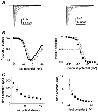
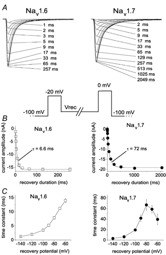
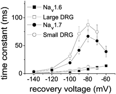
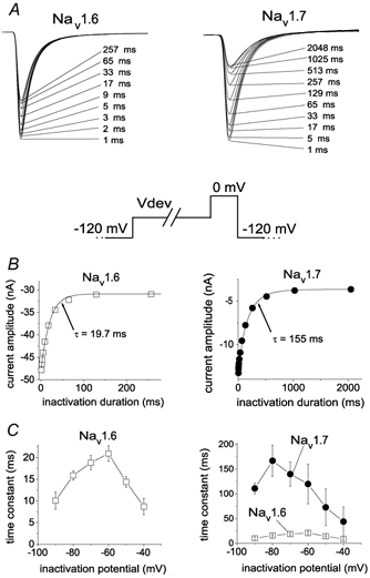
While large, myelinated dorsal root ganglion (DRG) neurons are capable of firing at high frequencies, small unmyelinated DRG neurons typically display much lower maximum firing frequencies. However, the molecular basis for this difference has not been delineated. Because the sodium currents in large DRG neurons exhibit rapid repriming (recovery from inactivation) kinetics and the sodium currents in small DRG neurons exhibit predominantly slow repriming kinetics, it has been proposed that differences in sodium channels might contribute to the determination of repetitive firing properties in DRG neurons. A recent study demonstrated that Nav1.7 expression is negatively correlated with conduction velocity and DRG cell size, while the Nav1.6 voltage-gated sodium channel has been implicated as the predominant isoform present at nodes of Ranvier of myelinated fibres. Therefore we characterized and compared the functional properties, including repriming, of recombinant Nav1.6 and Nav1.7 channels expressed in mouse DRG neurons. Both Nav1.6 and Nav1.7 channels generated fast-activating and fast-inactivating currents. However recovery from inactivation was significantly faster (approximately 5-fold at -70 mV) for Nav1.6 currents than for Nav1.7 currents. The recovery from inactivation of Nav1.6 channels was also much faster than that of native tetrodotoxin-sensitive sodium currents recorded from small spinal sensory neurons, but similar to that of tetrodotoxin-sensitive sodium currents recorded from large spinal sensory neurons. Development of closed-state inactivation was also much faster for Nav1.6 currents than for Nav1.7 currents. Our results indicate that the firing properties of DRG neurons can be tuned by regulating expression of different sodium channel isoforms that have distinct repriming and closed-state inactivation kinetics.


Figures

Figure 1
Figure 1. Comparison of Nav1.6r and Nav1.7r current properties
Representative traces from Nav1.8-null DRG neurons expressing Nav1.6r channels (A) and Nav1.7r channels (B) are shown. The currents were elicited by 50 ms test pulses to various potentials from −80 to 40 mV. Cells were held at −120 mV. C, normalized peak current-voltage relationship for Nav1.6r (▪; n = 16) and Nav1.7r channels (•; n = 10). D, voltage dependence of Nav1.6r (▪; n = 16) and Nav1.7r (•; n = 12) sodium current steady-state inactivation. Steady-state inactivation was estimated by measuring the peak current amplitude elicited by 20 ms test pulses to 0 mV after 500 ms prepulses to potentials over the range of −130 mV to −10 mV. Current is plotted as a fraction of the maximum peak current. E, activation time constants as a function of voltage are shown for Nav1.6r (▪; n = 14) and Nav1.7r (•; n = 7) currents in Nav1.8-null DRG neurons. F, inactivation time constants are shown for Nav1.6r (▪; n = 14) and Nav1.7r (•; n = 7) currents in Nav1.8-null DRG neurons as a function of voltage. The activation and inactivation time constants were estimated from Hodgkin-Huxley m3h fits to the currents elicited by 50 ms step depolarizations to voltages ranging from −40 to +40 mV.
Figure 2
Figure 2. Repriming (recovery from inactivation) kinetics are much faster for Nav1.6r than Nav1.7r channels in Nav1.8-null DRG neurons
A, family of current traces from representative neurons expressing Nav1.6r channels (left) and Nav1.7r channels (right) showing the rate of recovery from inactivation at −80 mV. The standard repriming voltage protocol is shown below. The cells were prepulsed to −20 mV for 20 ms to inactivate all of the current, then brought back to the recovery potential (Vrec) for increasing recovery durations prior to the test pulse to 0 mV. The maximum pulse rate was 0.5 Hz. The times indicated for each trace shown in A correspond to the recovery duration for that trace. B, the time course for recovery from inactivation of peak Nav1.6r (left) and Nav1.7r (right) currents in A are shown (note different time scales on x-axes). The continuous curve is a single exponential function fitted to the data, with a time constant τ of 6.6 ms for the Nav1.6r currents and 72 ms for the Nav1.7r currents. C, the time constants for recovery from inactivation of Nav1.6r (left) and Nav1.7r (right) currents are shown plotted as a function of voltage (note different time scales on y-axes). Time constants were estimated from single exponential fits to time courses measured at recovery potentials ranging from −140 to −60 mV with the protocol shown in A for currents recorded from neurons transfected with Nav1.6r channels (n = 15) or Nav1.7r channels (n = 11).
Figure 3
Figure 3
The recovery time constants for Nav1.6r (▪) and Nav1.7r channels (•) expressed in Nav1.8-null DRG neurons are compared to those for TTX-sensitive currents in large DRG neurons (▪; from Everill et al. 2001) and TTX-sensitive currents in small DRG neurons (○; from Cummins et al. 1998).
Figure 4
Figure 4. Recovery time constants for Nav1.6r channels are not altered by changes in the inactivating prepulse
A, the three different inactivating prepulses used to examine the repriming kinetics of Nav1.6r channels are shown. B, the repriming time constants measured with the different protocols for Nav1.6r channels are shown. The first protocol used a 20 ms prepulse to −20 mV (▪), the second protocol used a 20 ms prepulse to +20 mV (•) and the third protocol used a 50 ms prespulse to −20 mV (▴) to inactivate the Nav1.6r channels. The repriming kinetics were always fast for Nav1.6r channels and were not altered by the use of different inactivating prepulses.
Figure 5
Figure 5. Development of closed-state inactivation is more rapid for Nav1.6r than for Nav1.7r channels expressed in Nav1.8-null DRG neurons
A, family of current traces showing the rate of development of inactivation for Nav1.6r channels (left) and Nav1.7r channels (right) at −70 mV. The standard development of inactivation voltage protocol is shown below. From a holding potential of −120 mV, the cells were prepulsed to −70 mV (Vdev) for increasing durations, then stepped to 0 mV to determine the fraction of current inactivated during the prepulse. The duration of the inactivation prepulse for each data trace is indicated. B, time course for development of inactivation for the peak currents for Nav1.6r and Nav1.7r in A are shown. The continuous curve is a single exponential function fitted to the data, with a time constant of 19.7 ms for Nav1.6r channels (left) and 155 ms for Nav1.7r channels (right). C, the time constants for development of inactivation are shown plotted as a function of voltage. Time constants were estimated from single exponential fits to time courses measured at recovery potentials ranging from −90 to −40 mV with the protocol shown in A for currents recorded from neurons transfected with Nav1.6r channels (▪, left and right; n = 14) and neurons transfected with Nav1.7r channels (•, right; n = 11). As can be seen in the right panel, the development of inactivation time constants for Nav1.6r currents was much faster than for Nav1.7 currents.
Figure 6
Figure 6. Ramp currents generated by Nav1.6r and Nav1.7r channels
A, representative current elicited in a Nav1.8-null DRG neuron expressing Nav1.6r channels by a 500 ms ramp depolarization from −100 to +30 mV is shown. The peak sodium current amplitude elicited in this cell with step depolarizations was 65.1 nA. B, representative current elicited in a Nav1.8-null DRG neuron expressing Nav1.7r channels by a 500 ms ramp depolarization from −100 to +30 mV is shown. The peak sodium current amplitude elicited in this cell with step depolarizations was 75.4 nA. C, The relative ramp current amplitude was significantly larger in cells expressing Nav1.7r channels (grey bar) than in cells expressing Nav1.6r channels (open bar).

Similar articles

Cited by

References

    1. Akopian AN, Sivilotti L, Wood JN. A tetrodotoxin-resistant voltage-gated sodium channel expressed by sensory neurons. Nature. 1996;379:257–262. - PubMed
    1. Akopian AN, Souslova V, England S, Okuse K, Ogata N, Ure J, Smith A, Kerr BJ, McMahon SB, Boyce S, Hill R, Stanfa LC, Dickenson AH, Wood JN. The tetrodotoxin-resistant sodium channel Nav1. 8 has a specialized function in pain pathways. Nature Neurosci. 1999;2:541–548. - PubMed
    1. Black JA, Cummins TR, Plumpton C, Chen YH, Hormuzdiar W, Clare JJ, Waxman SG. Upregulation of a silent sodium channel after peripheral, but not central, nerve injury in DRG neurons. J Neurophysiol. 1999;82:2776–2785. - PubMed
    1. Black JA, Dib-Hajj S, McNabola K, Jeste S, Rizzo MA, Kocsis JD, Waxman SG. Spinal sensory neurons express multiple sodium channel α-subunit mRNAs. Brain Res Mol Brain Res. 1996;43:117–132. - PubMed
    1. Black JA, Waxman SG. Sodium channel expression: a dynamic process in neurons and non-neuronal cells. Dev Neurosci. 1996;18:139–52. - PubMed

Publication types

MeSH terms

LinkOut - more resources